. 2003 Feb 17;160(4):553-64.
doi: 10.1083/jcb.200211113.

The inner membrane protein Mdm33 controls mitochondrial morphology in yeast

Affiliations

The inner membrane protein Mdm33 controls mitochondrial morphology in yeast

Marlies Messerschmitt et al. J Cell Biol. .

Abstract

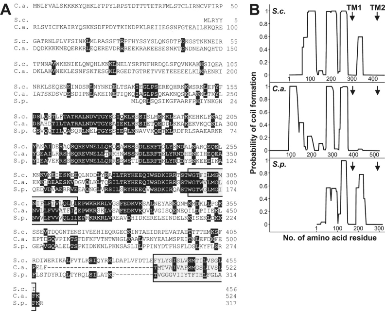
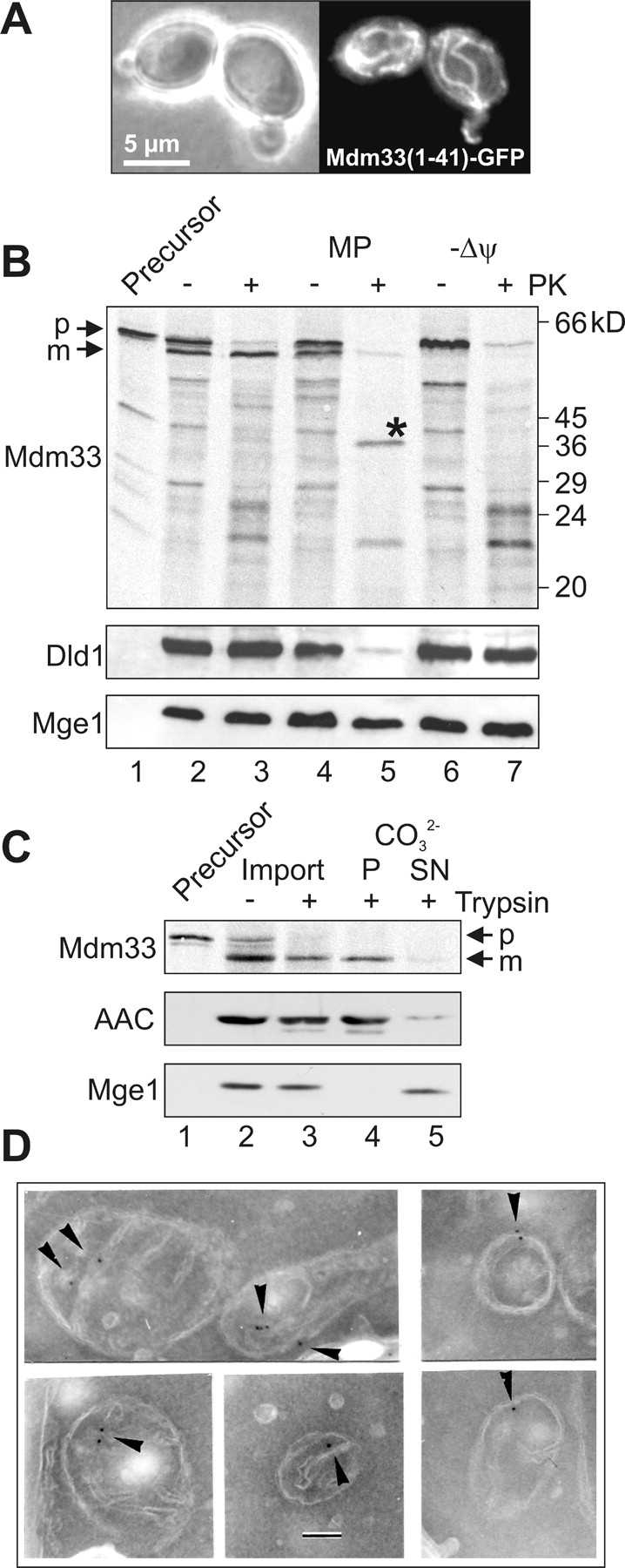
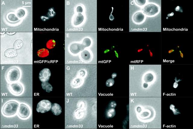
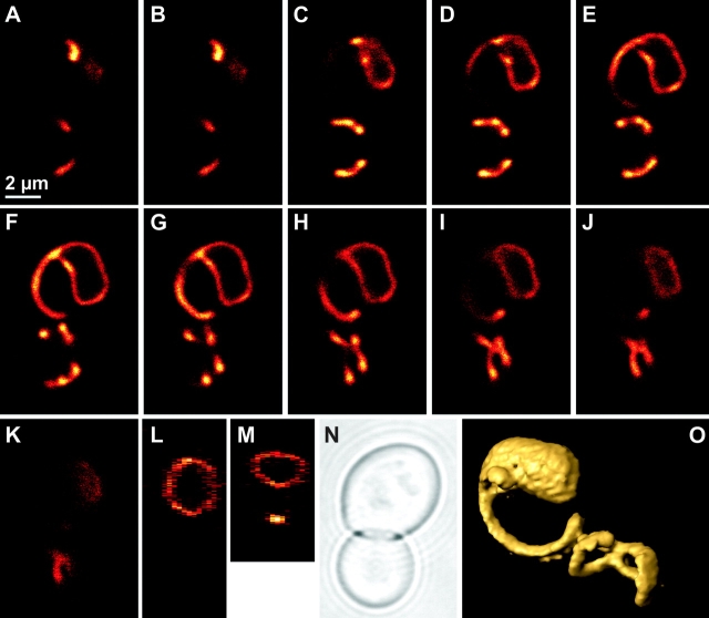
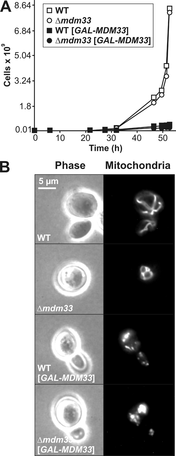
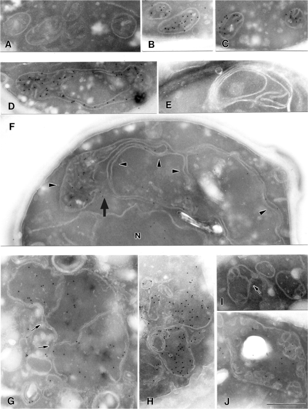
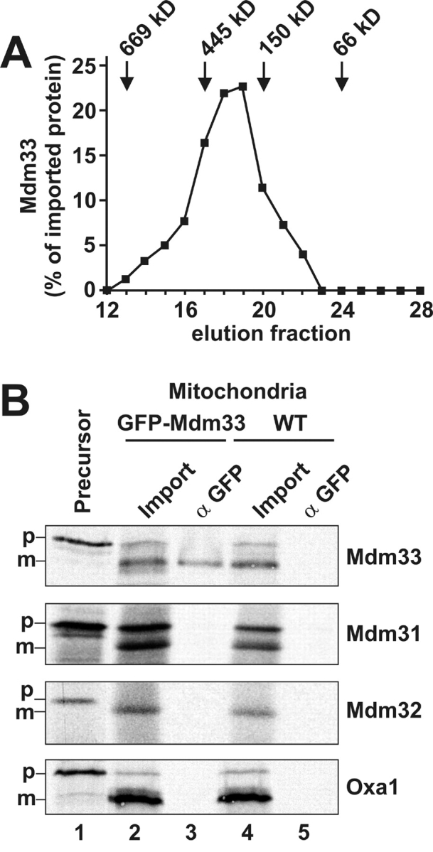
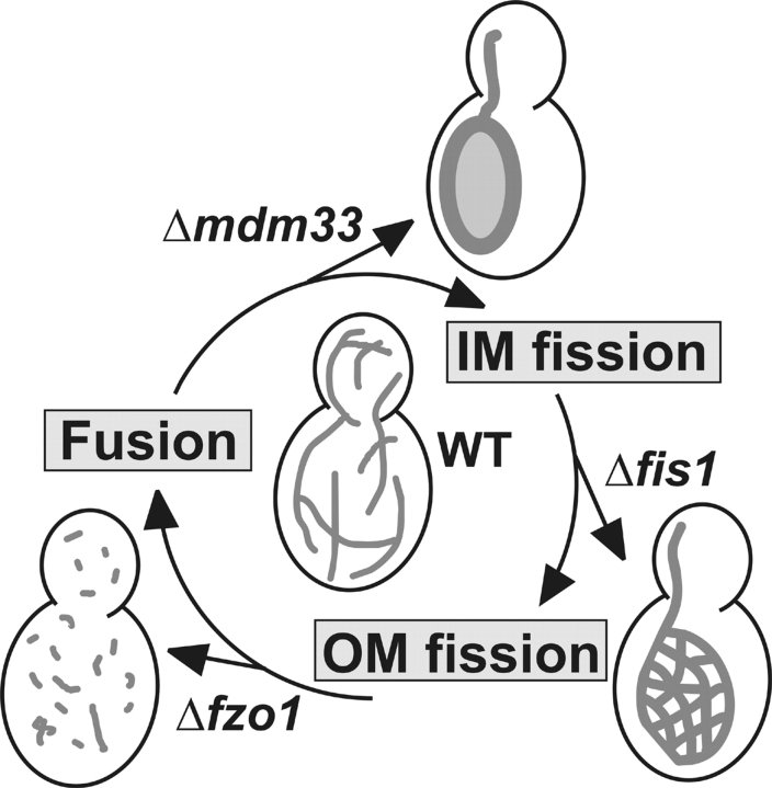
Mitochondrial distribution and morphology depend on MDM33, a Saccharomyces cerevisiae gene encoding a novel protein of the mitochondrial inner membrane. Cells lacking Mdm33 contain ring-shaped, mostly interconnected mitochondria, which are able to form large hollow spheres. On the ultrastructural level, these aberrant organelles display extremely elongated stretches of outer and inner membranes enclosing a very narrow matrix space. Dilated parts of Delta mdm33 mitochondria contain well-developed cristae. Overexpression of Mdm33 leads to growth arrest, aggregation of mitochondria, and generation of aberrant inner membrane structures, including septa, inner membrane fragments, and loss of inner membrane cristae. The MDM33 gene is required for the formation of net-like mitochondria in mutants lacking components of the outer membrane fission machinery, and mitochondrial fusion is required for the formation of extended ring-like mitochondria in cells lacking the MDM33 gene. The Mdm33 protein assembles into an oligomeric complex in the inner membrane where it performs homotypic protein-protein interactions. Our results indicate that Mdm33 plays a distinct role in the mitochondrial inner membrane to control mitochondrial morphology. We propose that Mdm33 is involved in fission of the mitochondrial inner membrane.


Figures

Figure 1.
Figure 1.
Mdm33 is a novel protein with predicted transmembrane segments and coiled-coil structures. (A) Comparison of the predicted amino acid sequences of Mdm33-related proteins. The amino acid sequences of Mdm33 of Saccharomyces cerevisiae (S.c.), CaShe9 of Candida albicans (C.a.), and SPAC823.13C of Schizosaccharomyces pombe (S.p.) were aligned using the DNAMAN software (Lynnon BioSoft). Amino acids that are identical in all three proteins are in black boxes, and gaps introduced to maximize the alignment are indicated by dashes. The positions of two predicted transmembrane segments (Hofmann and Stoffel, 1993), which are present in all three homologues, are indicated by gray boxes. (B) Plots of coiled-coil probabilities (Lupas et al., 1991) for the protein sequences shown in A. The window size used was 21. The positions of predicted transmembrane segments (TM1 and TM2) are indicated by arrows.
Figure 2.
Figure 2.
Mdm33 is a protein of the mitochondrial inner membrane. (A) Mitochondrial targeting of Mdm33(1–41)–GFP. Wild-type yeast cells expressing a chimeric protein consisting of the NH2-terminal 41 amino acid residues of Mdm33 fused to GFP (Mdm33[1–41]–GFP) were grown to log phase in YPGal (yeast extract-peptone-galactose) medium and examined by phase contrast (left panel) and fluorescence (right panel) microscopy. (B) Import in vitro of Mdm33. Mdm33 was synthesized in reticulocyte lysate in the presence of [35S]methionine (Precursor; lane 1) and imported into isolated mitochondria. The amount of precursor loaded in lane 1 corresponds to 10% of the amount used for each import reaction. For import in vitro, the protein was incubated with isolated mitochondria, and the organelles were reisolated by centrifugation. Equal aliquots were either left untreated on ice (lane 2), treated with proteinase K (PK; lane 3), converted to mitoplasts (MP) by hypotonic swelling in the absence (lane 4) or presence (lane 5) of PK, or import was performed in the presence of valinomycin to dissipate the membrane potential (Δψ) without (lane 6) and with (lane 7) subsequent treatment with PK. The asterisk indicates a specific fragment of imported Mdm33 that is generated only upon protease treatment of mitoplasts. The intactness of the mitochondrial membranes was controlled by immunoblotting using antisera against Dld1, an integral protein of the inner membrane that exposes its major part to the intermembrane space, and Mge1, a soluble matrix protein. p, precursor; m, mature form of Mdm33. (C) Carbonate extraction of imported Mdm33. Mdm33 was imported into mitochondria as in B. Aliquots were either left untreated on ice (lane 2) or treated with trypsin (lane 3). Then, protease treatment was stopped, and trypsin-treated samples were extracted with carbonate (CO3 2-) and separated into pellet (P; lane 4) and supernatant (SN; lane 5) fractions. All samples were precipitated with TCA and analyzed by SDS-PAGE and autoradiography. The fractionation into soluble and membrane proteins was controlled by immunoblotting using antisera against the ADP/ATP carrier (AAC), an integral protein of the inner membrane, and Mge1. p, precursor; m, mature form of Mdm33. (D) Immunocytochemistry of GFP–Mdm33. Yeast cells expressing a chimeric protein consisting of Mdm33 and GFP, inserted between the presequence and the mature part of Mdm33, were analyzed by immunoelectron microscopy using antibodies against GFP. Arrowheads point to inner membrane regions containing GFP–Mdm33. Bar, 100 nm.
Figure 3.
Figure 3.
Δmdm33 cells exhibit grossly altered mitochondrial morphology. (A–C) Mitochondrial morphology. Wild-type (WT) and Δmdm33 cells expressing mtGFP were grown to log phase in YPD medium and examined by phase contrast (left) and fluorescence (right) microscopy. (B) A representative Δmdm33 cell with a large ring-like mitochondrion. (C) A representative Δmdm33 cell with small interconnected ring-like mitochondria. (D) Double staining of mitochondria and cytosol. Δmdm33 cells coexpressing mtGFP and cRFP were grown to log phase in YPD medium and examined by confocal fluorescence microscopy. Left, bright field image taken in the transmission mode; right, merged confocal green and red fluorescence images. (E) Mitochondrial fusion. Δmdm33 cells of opposite mating types containing mitochondria preloaded with mtGFP or mtRFP were mated and analyzed by fluorescence microscopy. The distribution of mtGFP and mtRFP in a representative zygote containing a medial bud (asterisk) is shown. (F and I) Morphology of the ER. Wild-type and Δmdm33 cells expressing ER-targeted GFP were grown to log phase in glucose-containing synthetic minimal medium lacking methionine and examined by phase contrast and fluorescence microscopy. (G and J) Vacuolar morphology. Wild-type and Δmdm33 cells were grown to log phase in YPD medium, stained with 7-amino-4-chloromethylcoumarin, l-arginine amide to label vacuoles and examined by phase contrast and fluorescence microscopy. (H and K) Structure of the actin cytoskeleton. Wild-type and Δmdm33 cells were grown to log phase in YPD medium, fixed, stained with rhodamine-phalloidin to visualize filamentous actin, and examined by phase contrast and fluorescence microscopy.
Figure 4.
Figure 4.
Mitochondrial sphere in a Δmdm33 mutant cell. Δmdm33 cells expressing mtGFP were grown to log phase in YPGal medium. A representative cell was analyzed by confocal fluorescence microscopy. (A–K) Single optical planes (x/y); distance between serial planes is 285 nm. (L) Section corresponding to a z/y plane. (M) Section corresponding to a z/x plane. (N) Bright field image taken in the transmission mode. (O) Surface-rendered three-dimensional representation. This image has been turned 90° counter clockwise when compared with panels A–K.
Figure 5.
Figure 5.
Overexpression of Mdm33 induces growth arrest and aggregation of mitochondria. (A) Wild-type (WT) and Δmdm33 cells harboring empty vectors (open symbols) or multicopy plasmids with the MDM33 coding sequence under control of the GAL1 promoter (filled symbols; GALMDM33) were grown in galactose-containing synthetic complete medium lacking histidine to select for the overexpressing plasmid. At the indicated time points, the OD578 was measured, cultures were diluted when the OD exceeded 1, and growth was plotted as total number of cells. (B) Wild-type and Δmdm33 cells harboring empty vectors or multicopy plasmids with the MDM33 coding sequence under control of the GAL1 promoter (GALMDM33) were grown overnight in galactose-containing synthetic complete medium lacking histidine and examined by phase contrast (left) and fluorescence (right) microscopy. Mitochondria were visualized with mtGFP.
Figure 6.
Figure 6.
Ultrastructure of mitochondria in Mdm33-deficient and Mdm33-overexpressing cells. (A-C) Cross sections of mitochondria of wild-type cells. (D-F) Cross sections of mitochondria of Δmdm33 cells. N, nucleus. Arrowheads point to extended interconnected mitochondrial structures; the arrow points to a putative intramitochondrial fusion site. (G–J) Cross sections of mitochondria of Mdm33-overexpressing cells. Induction of GALMDM33 was as described for Fig. 5 B. Arrows point to inner membrane septa. (B, C, D, F, G, H and J) Immunogold labeling of mtGFP to identify the mitochondrial matrix compartment. It should be noted that the mitochondrial outer membrane appears relatively faint due to its low protein content. Bar, 500 nm.
Figure 7.
Figure 7.
Mdm33 assembles into higher molecular complexes. (A) Gel filtration of Mdm33. Mdm33 protein synthesized in vitro was imported into wild-type mitochondria as in Fig. 2 B. After completion of import, mitochondria (1 mg) were washed, reisolated, and solubilized in 300 μl buffer containing 0.5% Triton X-100. Nonsolubilized and aggregated material was separated by centrifugation, and the supernatant was loaded on a superose-6 column. After chromatography, the collected fractions were analyzed by SDS-PAGE, blotting to nitrocellulose, and autoradiography. Recovered Mdm33 was quantified by densitometry. Marker proteins thyroglobulin, apoferritin, alcohol dehydrogenase, and bovine serum albumin were used to calibrate the column. (B) Coimmunoprecipitation of Mdm33. Mdm33 translated in vitro was imported into mitochondria harboring GFP–Mdm33 or nontagged Mdm33 (WT). Mitochondria were washed, reisolated, lysed with Triton X-100, and subjected to coimmunoprecipitation with antibodies directed against GFP. In control reactions, two other putative inner membrane proteins involved in mitochondrial morphogenesis, Mdm31 and Mdm32, and a nonrelated inner membrane protein, Oxa1, were imported. Lane 1, [35S]methionine-labeled precursor proteins, corresponding to 10% of the amounts added to the import reactions. Lane 2, import reactions using GFP–Mdm33 mitochondria, corresponding to 10% of the amounts used for the coimmunoprecipitations. Lane 3, coimmunoprecipitations from GFP–Mdm33 mitochondria using anti-GFP antiserum. Lane 4, import reactions using wild-type mitochondria. Lane 5, coimmunoprecipitations from wild-type mitochondria using anti-GFP antiserum. p, precursor forms; m, mature forms of imported proteins.
Figure 8.
Figure 8.
Hypothetical cycle of epistatic relationships between components with a role in mitochondrial dynamics. Arrows leaving the cycle indicate the mitochondrial phenotypes of the deletion mutants. Each gene deletion is epistatic to the following one, i.e., double mutants obtained by genetic crosses display the phenotype of the mutant that is preceding in the cycle. See text for further details. IM, inner membrane; OM, outer membrane; WT, wild type.

Similar articles

Cited by

References

    1. Amberg, D.C. 1998. Three-dimensional imaging of the yeast actin cytoskeleton through the budding cell cycle. Mol. Biol. Cell. 9:3259–3262. - PMC - PubMed
    1. Andaluz, E., J.J. Coque, R. Cueva, and G. Larriba. 2001. Sequencing of a 4.3 kbp region of chromosome 2 of Candida albicans reveals the presence of homologues of SHE9 from Saccharomyces cerevisiae and of bacterial phosphatidylinositol-phospholipase C. Yeast. 18:711–721. - PubMed
    1. Bereiter-Hahn, J. 1990. Behavior of mitochondria in the living cell. Int. Rev. Cytol. 122:1–63. - PubMed
    1. Bleazard, W., J.M. McCaffery, E.J. King, S. Bale, A. Mozdy, Q. Tieu, J. Nunnari, and J.M. Shaw. 1999. The dynamin-related GTPase Dnm1 regulates mitochondrial fission in yeast. Nat. Cell Biol. 1:298–304. - PMC - PubMed
    1. Boldogh, I.R., H.-C. Yang, and L.A. Pon. 2001. Mitochondrial inheritance in budding yeast. Traffic. 2:368–374. - PubMed

Publication types

MeSH terms

Associated data