. 2003 Feb 17;160(4):597-604.
doi: 10.1083/jcb.200210053.

Amassin, an olfactomedin protein, mediates the massive intercellular adhesion of sea urchin coelomocytes

Affiliations

Amassin, an olfactomedin protein, mediates the massive intercellular adhesion of sea urchin coelomocytes

Brian J Hillier et al. J Cell Biol. .

Abstract

Sea urchins have a fluid-filled body cavity, the coelom, containing four types of immunocytes called coelomocytes. Within minutes after coelomic fluid is removed from the body cavity, a massive cell-cell adhesion of coelomocytes occurs. This event is referred to as clotting. Clotting is thought to be a defense mechanism against loss of coelomic fluid if the body wall is punctured, and it may also function in the cellular encapsulation of foreign material and microbes. Here we show that this intercoelomocyte adhesion is mediated by amassin, a coelomic plasma protein with a relative molecular mass (Mr) of 75 kD. Amassin forms large disulfide-bonded aggregates that adhere coelomocytes to each other. One half of the amassin protein comprises an olfactomedin (OLF) domain. Structural predictions show that amassin and other OLF domain-containing vertebrate proteins share a common architecture. This suggests that other proteins of the OLF family may function in intercellular adhesion. These findings are the first to demonstrate a function for a protein of the OLF family.


Figures

Figure 1.
Figure 1.
The clotting of coelomocytes. Sea urchin coelomocytes form a massive cellular clot by rapid intercellular adhesion. The contents of the coelomic cavity of one animal was poured into a beaker, constantly swirled, and top view photographs were taken. (A) After 10 s, aggregates of coelomocytes cannot be seen. (B) After 50 s, the cells have begun to form aggregations. (C) After 70 s, the clot begins to form with stringy cellular masses continuing to adhere to each other. (D) By 150 s, clot formation is complete. Bar, 1 cm.
Figure 2.
Figure 2.
An assay for the level of coelomocyte clotting. The degree of coelomocyte clotting depends on the concentration of CFP. Complete clotting occurs at 3 μg CFP/ml. Data points are the mean of five experiments with the same batch of cells (standard error bars shown). Clot completion is defined as the level of clotting that 10 μg CFP would promote compared with no clotting with buffer alone.
Figure 3.
Figure 3.
DEAE-Sepharose chromatography of cell-free coelomic plasma. A silver-stained 7.5% SDS-PAGE separation of proteins eluting from the DEAE column upon application of a linear salt gradient that increases left to right. Numbers on top of lanes represent individual fractions. A 10-μl aliquot of each fraction was subjected to the clotting assay; + denotes the ability to clot, and − fractions had no detectable clotting activity. The positive fractions are highly enriched for a 75-kD protein called amassin. S, molecular mass standards presented in kD.
Figure 4.
Figure 4.
Antibodies to amassin prevent clotting. (A) Western blots of CFP show that the antibody reacts only with the 75 kD amassin. CFP (5 μg) was loaded onto lane 1 and stained with Coomassie. The sample for lane 2 was 300 ng CFP and detected by Western blot with antiamassin. (B) Preincubation of CFP with antiamassin inhibits coelomocyte clotting. Crude antiserum was incubated with sufficient CFP to induce complete clotting (3 μg/ml) for 15 min on ice in a volume of 20 μl before adding to washed coelomocytes in the clotting assay.
Figure 5.
Figure 5.
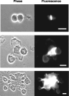
Antibody localizes amassin between adhering coelomocytes. The left panels are phase contrast, and the right panels are fluorescence with antiamassin. In the top set, amassin binds two coelomocytes together. In the bottom two sets, larger groups of coelomocytes are shown. Bars, 15 μm.
Figure 6.
Figure 6.
Amassin is disulfide bonded into large aggregates. Samples of CFP were incubated with decreasing concentrations of DTT (twofold dilutions starting from 100 mM in lane 1 to 24 μM in lane 13; lane 14 had no DTT) in SDS-PAGE gel loading buffer before separation on a 7.5% gel and immunoblotted with antiamassin. If disulfide bond-reducing agents are excluded from the sample buffer, amassin does not enter the 5% stacking gel (lane 14). Complete reduction of disulfides in lane 1 shows the 75-kD reacting band. The graded concentration series of DTT displays what can be classified as monomers, dimers, and oligomers of amassin.
Figure 7.
Figure 7.
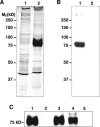
Amassin is released as coelomocyte clots are dissociated into single cells. (A) Washed clots were dissociated in DTT, and the 10,000-g supernatant was recovered (lane 2). A control sample, lane 1 is the supernatant released from the same DTT treatment to washed, clot-inhibited coelomocytes run on a 10% silver-stained SDS-PAGE gel. DTT dissociates the clot into single cells and releases amassin. (B) An immunoblot probed with antiamassin. Amassin is present in clots of coelomocytes (lane 1), but almost undetectable levels (only after long exposures) are present in a sample of washed clot-inhibited coelomocytes (lane 2). (C) Samples of CFP (300 ng per lane, lane 1 no treatment) were subjected to ultracentrifugation either in the absence (lanes 2–3) or presence (lanes 4–5) of 10 mM DTT. The resulting supernatants (lanes 2 and 4) and pellets (lanes 3 and 5) detected by immunoblot with antiamassin show that amassin is present as a large, disulfide-bonded aggregate which can be dissociated with DTT.
Figure 8.
Figure 8.
Confirmation of amassin cloning by recombinant protein reactivity and relative molecular mass agreement. (A) The bacterial expression of the derived amino acid sequence from the amassin OLF domain (aa 228–495) is recognized by the antibodies to native amassin. Escherichia coli lysates of preinduced (lane 1) and 30 min postinduction (lane 2) are shown stained with Coomassie; lanes 3 and 4 are the same samples but immunoblotted with antiamassin, which specifically detects the OLF domain of the expected size. (B) Amassin is heavily glycosylated. Purified amassin was digested with PNGase-F to cleave off the N-linked oligosaccharides. SDS-PAGE analysis of the digest (lane 2) showed a decrease in M r relative to the minus enzyme control (lane 1), from 75 kD to ∼60 kD.
Figure 9.
Figure 9.
Analysis of the amassin sequence. Sequence alignment of five OLF family members in only the OLF domains (A) or only the NH2-terminal β-regions (B). AMAS, amassin; NOEL, noelin-1; MYOC, myocilin; TIAR, tiarin; OLFM, olfactomedin. Amino acid identities are dark shaded, similarities are light shaded, and boxes enclose both. Positions which contain cysteines are noted by an asterisk above the alignment. Predicted secondary structural motifs are depicted with arrows for β-strands and a cylinder for an α-helix. (C) Comparison of key structural motifs in five full-length OLF family members. From NH2 to COOH terminus there is a signal sequence (1), a short β-region (2), a long coiled-coil motif (3), and the OLF domain (4). All regions containing cysteines are shown. Sequence data for amassin is available from GenBank/EMBL/DDBJ under accession no. AF533649.

References

    1. Barembaum, M., T.A. Moreno, C. LaBonne, J. Sechrist, and M. Bronner-Fraser. 2000. Noelin-1 is a secreted glycoprotein involved in generation of the neural crest. Nat. Cell Biol. 2:219–225. - PubMed
    1. Bateman, A., E. Birney, L. Cerruti, R. Durbin, L. Etwiller, S.R. Eddy, S. Griffiths-Jones, K.L. Howe, M. Marshall, and E.L. Sonnhammer. 2002. The Pfam protein families database. Nucleic Acids Res. 30:276–280. - PMC - PubMed
    1. Bertheussen, K., and R. Seijelid. 1978. Echinoid phagocytes in vitro. Exp. Cell Res. 111:401–412. - PubMed
    1. Bookhout, C.G., and N.D. Greenburg. 1940. Cell types and clotting reactions in the echinoid, Mellita quinquiesperforata Biol. Bull. 79:309–320.
    1. Boolootian, R.A., and A.C. Giese. 1959. Clotting of echinoderm coelomic fluid. J. Exp. Zool. 140:207–229. - PubMed

Publication types

Associated data