. 2003 Mar 4;100(5):2409-14.
doi: 10.1073/pnas.0437660100. Epub 2003 Feb 18.

Oligomeric and polymeric aggregates formed by proteins containing expanded polyglutamine

Affiliations

Oligomeric and polymeric aggregates formed by proteins containing expanded polyglutamine

S Iuchi et al. Proc Natl Acad Sci U S A. .

Abstract

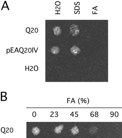
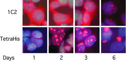
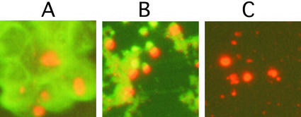
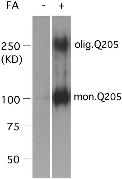
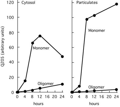
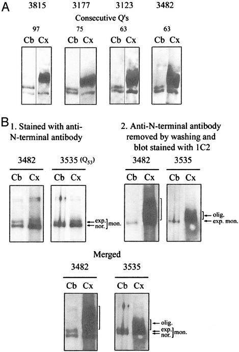
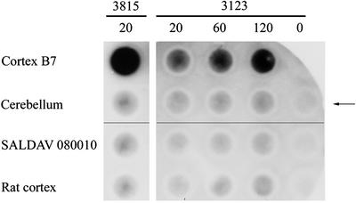
Neurological diseases resulting from proteins containing expanded polyglutamine (polyQ) are characteristically associated with insoluble neuronal inclusions, usually intranuclear, and neuronal death. We describe here oligomeric and polymeric aggregates formed in cells by expanded polyQ. These aggregates are not dissociated by concentrated formic acid, an extremely effective solvent for otherwise insoluble proteins. Perinuclear inclusions formed in cultured cells by expanded polyQ can be completely dissolved in concentrated formic acid, but a soluble protein oligomer containing the expanded polyQ and released by the formic acid is not dissociated to monomer. In Huntington's disease, a formic acid-resistant oligomer is present in cerebral cortex, but not in cerebellum. Cortical nuclei contain a polymeric aggregate of expanded polyQ that is insoluble in formic acid, does not enter polyacrylamide gels, but is retained on filters. This finding shows that the process of polymerization is more advanced in the cerebral cortex than in cultured cells. The resistance of oligomer and polymer to formic acid suggests the participation of covalent bonds in their stabilization.


Figures

Figure 1
Figure 1
Solubility of Q20 peptides in formic acid. Q20 and pEA Q20 IV were added to water and a solution of 2% SDS in an amount sufficient to make a 2-mM solution. The suspensions were centrifuged at 14,000 rpm for 5 min in an Eppendorf centrifuge. Any sediment was resuspended in water, and an aliquot was allowed to evaporate on a glass plate. The residue on the plate was photographed against a black background with overhead illumination. (A) Q20 and pEA Q20 IV were largely insoluble in water or in a solution of SDS. When 90% formic acid (FA) was substituted as solvent, both peptides dissolved, leaving no sediment. (B) Solubility of Q20 in different concentrations of formic acid (FA).
Figure 2
Figure 2
Appearance and number of inclusions after induction of PC12-Q205 cells. Expression of the construct encoding Q205, flanked by 67 N-terminal and 17 C-terminal residues, followed by 6xHis, was induced from an ecdysone-inducible promotor, using ponasterone A. The 1C2 antibody, which is known to stain inclusions poorly, showed mainly diffuse cytoplasmic staining, declining with time, whereas staining by anti-tetra-His showed mainly inclusions, beginning as early as day 1, first increasing in number with time but later only in size, the number then being reduced.
Figure 3
Figure 3
Purification of inclusions. Inclusions harvested 6 days after induction of PC12-Q205 cells were purified. The inclusions were stained with anti-c-Myc coupled indirectly to Cy3 (red) and nonspecific proteins were stained with anti-rabbit IgG-FITC (green). (A) No purification. (B) After extraction with guanidinium chloride. (C) After extraction with SDS/2-ME. (Magnifications: ×300.)
Figure 4
Figure 4
Synthesis by PC12-Q205 cells of an oligomer containing expanded polyQ. Purified inclusions, with or without formic acid (FA) treatment, were boiled in SDS/2-ME and submitted to electrophoresis through 7.5% polyacrylamide. After transfer to nitrocellulose, the proteins were stained with the antibody 1C2. In the absence of formic acid pretreatment, neither monomer nor oligomer was detected. After pretreatment with 90% formic acid for 1 h at 37°C, electrophoresis revealed a strong band of monomer corresponding to an apparent molecular mass of ≈100 kDa; the true molecular mass should be 36,340, but expanded polyQ reduces mobility in gel electrophoresis. In addition to the monomer, an oligomer with a considerably higher molecular mass is clearly stained by the 1C2 antibody. This film is overexposed to demonstrate the oligomer, but estimation of staining intensity by IR fluorescence showed that the oligomer was present in a range from 0.5% to 3% of monomer. The ratio of the mobility of the oligomer to that of the monomer was 0.21.
Figure 5
Figure 5
Kinetics of appearance of monomer and oligomer in cytosol and particulates of induced PC12-Q205 cells. One day after inoculation, PC12-Q205 cells were induced by 10 μM ponasterone A and harvested by trypsinization after 0, 4, 8, 12, and 24 h of induction. Sonicated extracts were fractionated into soluble and particulate fractions (containing the inclusions) and both fractions were analyzed for Q205 by Western blotting. The amount of Q205 determined by IR fluorescence is shown per 5 μg of cellular protein. Monomer and oligomer were readily detected in both cytosol and particulates within 4 h of induction. The amount of monomer always exceeded the amount of oligomer, but the particulates account for an appreciable part of the total oligomer at the earliest time points (see text).
Figure 6
Figure 6
Oligomeric state of soluble expanded huntingtin in cerebral cortex in Huntington's disease. (A) Crude extracts of cortex and cerebellum were treated with 96% formic acid. Western blots prepared after electrophoresis were stained consecutively with anti-N-terminal antibody (30) and 1C2. All specimens of cortex produced a broad oligomeric band of expanded huntingtin absent from the cerebellum. In contrast, all of the cerebellar specimens, but none of the cortical specimens, produced a clear band of monomeric expanded huntingtin. (B) Blots prepared after electrophoresis of formic acid-treated crude brain extracts were photographed after staining with the N-terminal antibody and again after staining with the 1C2 antibody. The expanded monomer, but not the oligomer, is clearly stained by the N-terminal antibody. The broad band of oligomer in the merged figure clearly extends above the corresponding monomer.
Figure 7
Figure 7
Polymeric state of expanded huntingtin in cerebral cortex in Huntington's disease. Purified nuclei of cortex and cerebellum of brain 3815 and of controls were extracted with 96% formic acid. The suspension was diluted with a 10-fold excess of Tris/SDS/2-ME, boiled for 5 min, and immediately passed through cellulose acetate filters. The retained material was visualized by chemiluminescence by using antibody 1C2 and the ECL plus kit. Control SALDAV 080010 was a lymphoblast line possessing a huntingtin with 79 glutamine residues. Rat cortex, whose huntingtin possesses Q8 (51), should not be stainable by 1C2. Cortex 3815: 20,000 nuclei gave a strong signal. The corresponding cerebellum, SALDAV 080010, and rat cortex gave weak signals. Cortex 3123: 120,000 nuclei produced a weaker signal than 20,000 nuclei of cortex 3815. Arrow indicates control without nuclei. Because chemiluminescence does not provide a quantitative measure of the amount of expanded polyQ, quantitation was performed by using IR fluorescence (see Table 1).

References

    1. La Spada A R, Wilson E M, Lubahn D B, Harding A E, Fischbeck K H. Nature. 1991;352:77–79. - PubMed
    1. The Huntington's Disease Collaborative Research Group. Cell. 1993;72:971–983. - PubMed
    1. Gusella J F, MacDonald M E. Nat Rev Neurosci. 2000;1:109–115. - PubMed
    1. Perutz M F, Johnson T, Suzuki M, Finch J T. Proc Natl Acad Sci USA. 1994;91:5355–5358. - PMC - PubMed
    1. Eckert R L, Green H. Cell. 1986;46:583–589. - PubMed

Publication types

MeSH terms

LinkOut - more resources