. 2003 Mar;9(3):280-6.
doi: 10.1261/rna.2173903.

Recurring features of local tertiary structural elements in RNA molecules exemplified by hepatitis D virus RNA

Affiliations

Recurring features of local tertiary structural elements in RNA molecules exemplified by hepatitis D virus RNA

David A Circle et al. RNA. 2003 Mar.

Abstract

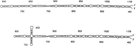
Elements of local tertiary structure in RNA molecules are important in understanding structure-function relationships. The loop E motif, first identified in several eukaryotic RNAs at functional sites which share an exceptional propensity for UV crosslinking between specific bases, was subsequently shown to have a characteristic tertiary structure. Common sequences and secondary structures have allowed other examples of the E-loop motif to be recognized in a number of RNAs at sites of protein binding or other biological function. We would like to know if more elements of local tertiary structure, in addition to the E-loop, can be identified by such common features. The highly structured circular RNA genome of the hepatitis D virus (HDV) provides an ideal test molecule because it has extensive internal structure, a UV-crosslinkable tertiary element, and specific sites for functional interactions with proteins including host PKR. We have now found a UV-crosslinkable element of local tertiary structure in antigenomic HDV RNA which, although differing from the E-loop, has a very similar pattern of sequence and secondary structure to the UV-crosslinkable element found in the genomic strand. Despite the fact that the two structures map close to one another, the sequences comprising them are not the templates for each other. Instead, the template regions for each element are additional sites for potential higher order structure on their respective complementary strands. This wealth of recurring sequences interspersed with base-paired stems provides a context to examine other RNA species for such features and their correlations with biological function.


Figures

FIGURE 1.
FIGURE 1.
Schematic diagram of the HDV genome. The collapsed rod structure with the numbering system proposed by Wang et al. (1986) is depicted. For genomic RNA, the 5′ direction is clockwise; whereas for antigenomic RNA, the 5′-to-3′ direction is counterclockwise. The highly conserved “Viroid-Like Region” and the less well-conserved “Protein Coding Region,” first proposed by Branch et al. (1989a), are shown separated by a heavy vertical line. Also shown are the open reading frame for the HDV antigen, a structural protein encoded by the antigenomic strand (“δ Antigen ORF”—unshaded box); the sequence comprising the antigenomic ribozyme (“δ antigenomic ribozyme”—filled box) and its cut site between residues 900 and 901 (“Cleavage Site” with downward pointing arrow); the region shown by Sharmeen et al. (1988) to influence the HDV antigenomic ribozyme activity (“δ Ribozyme Regulatory Element”—gray box); the cut site for the genomic ribozyme between residues 685 and 686 (“Cleavage Site”with upward pointing arrow); the boundaries of the 13/3 RNA transcript covering residues 482–963 (“δ 13/3 Transcript”—arrowheads); and the position at antigenomic residue A1012 that undergoes RNA editing (“Editing Site” with downward pointing arrow).
FIGURE 2.
FIGURE 2.
Comparison of the antigenomic (A) and genomic (B) HDV UV-sensitive structures. Note that the antigenomic sequence has been inverted with respect to the map in Figure 1 ▶ to facilitate comparison with its genomic counterpart. Both can be drawn as internal loops (which reflect the still unknown higher order folding that must be present, pending X-ray or NMR analysis), flanked by runs of Watson–Crick base pairs. Conserved nucleotides are indicated by upper case letters, whereas nonconserved bases are shown in lower case. The diagonal arrows indicate the bases that undergo UV-crosslinking.
FIGURE 3.
FIGURE 3.
Positions of HDV RNA blocs comprising tertiary structural elements and their templates depicted in both linear and collapsed rod maps. The numbering system of Wang et al. (1986) is employed, and the positions of the first (base 1) and last (base 1679) bases are indicated. The 5′-to-3′ directions are shown in each case. In all maps, the RNA blocs that undergo UV crosslinking in the strand depicted are shown as filled rectangles, whereas templates for such elements in the opposite strand are shown in rectangles hatched with diagonal lines. The “endpoint” bases of the collapsed rod, 793 and 1637, are indicated. The genomic ribozyme cleavage site is depicted by an “R” with an upward-pointing arrow. The antigenomic ribozyme cut site is indicated by an “R” with a downward-pointing arrow; the editing site, by an “E” with a downward-pointing arrow. Upper drawings: The genomic UV-crosslinkable element blocs (filled rectangles) comprise bases 710–735 and 857–875, and the template blocs for the antigenomic UV-crosslinkable element (hatched rectangles) include bases 686–705 and 878–899. Lower drawings: The antigenomic UV-crosslinkable element blocs (filled) are bases 686–705 and 878–899; the templates for the genomic element (hatched) include bases 710–735 and 857–875.
FIGURE 4.
FIGURE 4.
Recurring elements in HDV regions with potential tertiary structure. These drawings each represent a close-up of the critical parts of the collapsed rod maps shown in Figure 3 ▶. The antigenomic region is shown above; the genomic, below. Cleavage points for the antigenomic (“AG Rbz”) and genomic (“G Rbz”) ribozyme activities are indicated, as are 5′ and 3′ termini. The loops shown at the left-hand end of each structure symbolize the nearby “closed end” of the collapsed rod centered at base 793. The bases that undergo UV crosslinking (U701, G889—antigenomic strand; U712, U865—genomic strand) are starred. Potential base pairs (except those involving starred bases) are drawn according to Wang et al. (1986), although their existence as such within the regions of bold or highlighted type is unlikely. Upper drawing: The UV-crosslinkable element reported here is indicated, in the right half of the drawing, by the bold, raised (or lowered) type that highlights its recurring sequences. These blocs correspond to the filled rectangles in the HDV antigenomic map depicted in Figure 3 ▶. In like manner, the template sequences for the genomic UV-sensitive element are depicted, in the left half of the drawing, by italicized, highlighted open type (raised or lowered), which depicts the recurring sequences. These blocs correspond to the hatched rectangles in the Figure 3 ▶ genomic HDV map. Lower drawing: The previously mapped HDV genomic UV-crosslinkable element is indicated on the left via its recurring sequences (bold type, raised or lowered); the template for the antigenomic UV-crosslinkable element is similarly depicted on the right (italicized, highlighted open type, raised or lowered).
FIGURE 5.
FIGURE 5.
Alternate secondary structures for the HDV antigenomic strand. The UV-crosslinkable element of local tertiary structure is a common feature of both structures shown, represented by a “pinched” domain in the vicinity of residue 700 (lower strand). The alternate folding of bases between 700–750 and 800–850 is shown, forming a cruciform in the lower drawing. HDV genomic and antigenomic RNAs can both fold into two alternate configurations like the ones shown here, as first described by Branch and Robertson (1991). As proposed by Robertson et al. (1996), the two structures depicted here are likely to differ in their ability to bind host proteins such as PKR. In the antigenomic strand, the two alternate structures could regulate the relative affinities of PKR and the ADAR RNA editing enzyme.

Similar articles

Cited by

References

    1. Allison, L.A., Romaniuk, P.J., and Bakken, A.H. 1991. RNA–protein interactions of stored 5S RNA with TFIIIA and ribosomal protein L5 during Xenopus oogenesis. Dev. Biol. 144: 129–144. - PubMed
    1. Atmadja, J., Brimacombe, R., Blocker, H., and Frank, R. 1985. Investigation of the tertiary folding of Escherichia coli 16S RNA by in situ intra-RNA cross-linking within 30S ribosomal subunits. Nucleic Acids Res. 13: 6919–6936. - PMC - PubMed
    1. Barrell, B.J. 1971. Fractionation and sequence analysis of radioactive nucleotides. In Procedures in nucleic acids research (eds. G.L. Cantoni and D.R. Davies), Vol. 2, pp. 751–779. Harper & Row, New York.
    1. Bichko, V., Bank, S., and Taylor, J. 1997. Phosphorylation of the hepatitis delta virus antigens. J. Virol. 71: 512–518. - PMC - PubMed
    1. Branch, A.D. and Robertson, H.D. 1991. Efficient trans cleavage and a common structural motif for the ribozymes of the human HDV. Proc. Natl. Acad. Sci. 88: 10163–10167. - PMC - PubMed

Publication types

LinkOut - more resources