. 2003 Mar;9(3):331-45.
doi: 10.1261/rna.2144203.

Structure-function analysis of the 3' stem-loop of hepatitis C virus genomic RNA and its role in viral RNA replication

Affiliations

Structure-function analysis of the 3' stem-loop of hepatitis C virus genomic RNA and its role in viral RNA replication

Minkyung Yi et al. RNA. 2003 Mar.

Abstract

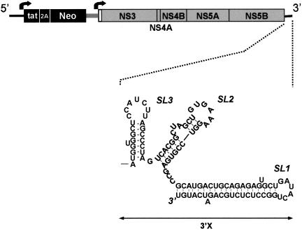
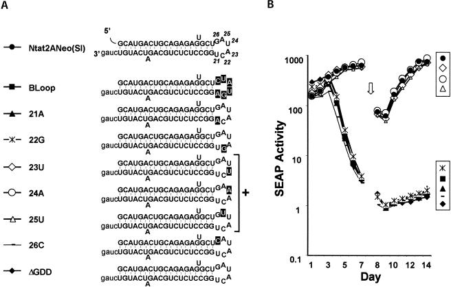
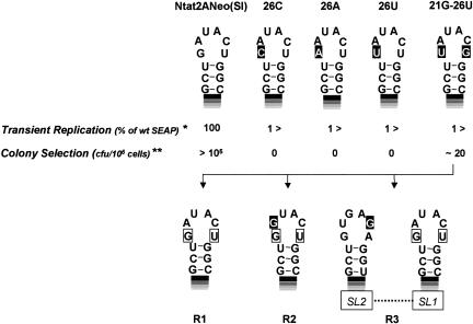
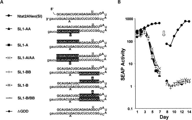
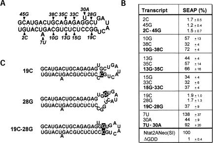
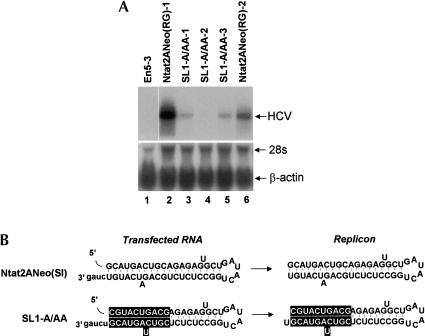
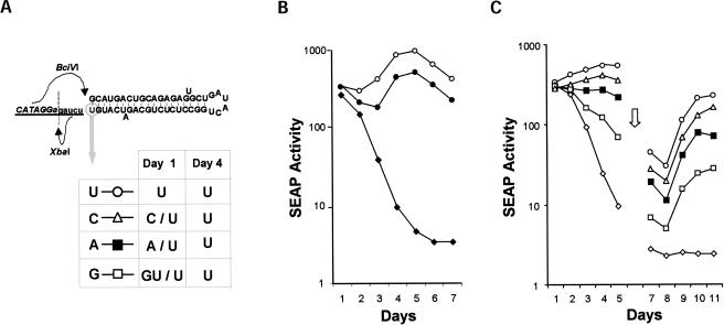
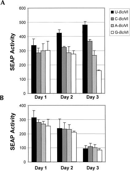
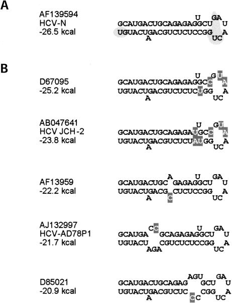
Previous studies indicate that the 3' terminal 46 nt of the RNA genome of hepatitis C virus (HCV) are highly conserved among different viral strains and essential for RNA replication. Here, we describe a mutational analysis of the 3' terminal hairpin (stem-loop I) that is putatively formed by this sequence and demonstrate its role in replication of the viral RNA. We show that single base substitutions within the 6-nt loop at positions adjacent to the stem abrogate replication of a subgenomic RNA, whereas substitutions in the three apical nucleotides were well tolerated without loss of replication competence. Single point mutations were also well tolerated within the middle section of the duplex, but not at the penultimate nucleotide positions near either end of the stem. However, complementary substitutions at the -19 and -28 positions (from the 3' end) restored replication competence, providing strong evidence for the existence of the structure and its involvement in RNA replication. This was confirmed by rescue of replicating RNAs from mutants containing complementary 10-nt block substitutions at the base of the stem. Each of these RNAs contained an additional U at the 3' terminus. Further experiments indicated a strong preference for U at the 3' terminal position (followed in order by C, A, and G), and a G at the -2 position. These features of stem-loop I are likely to facilitate recognition of the 3' end of the viral RNA by the viral RNA replicase.


Figures

FIGURE 1.
FIGURE 1.
A schematic depicting the organization of the dicistronic Ntat2ANeo(SI) replicon RNA (Yi et al. 2002) in which mutations were created within the 46 nt that putatively form a 3′ terminal stem-loop structure, SL1. An expanded view below the map of the replicon shows the predicted RNA secondary structure within the 3′X domain of the 3′NTR (Blight and Rice 1997).
FIGURE 2.
FIGURE 2.
Expression of SEAP by En5-3 cells following transient transfection with synthetic (filled circles) Ntat2ANeo(SI) RNA, and related replication-defective mutants, (filled diamonds) ΔGDD and (open triangle) ΔSL1, in the absence of G418 selection. Cell culture supernatant fluids were collected and replaced with fresh media at 24-h intervals. The harvested media was stored at 4°C until assayed for SEAP activity at the conclusion of the experiment. (A) Secretion of SEAP during successive 24-h intervals was normalized to that produced during the first 24 h after transfection (100%) for each culture. Declining SEAP secretion by cells transfected with ΔGDD and ΔSL1 is indicative of the failure of these transfected RNAs to replicate. (B) SEAP activities produced by cells transfected with Ntat2ANeo(SI) and the related ΔSL1 mutant, normalized as in A, are shown relative to that produced by cells transfected with the polymerase-defective ΔGDD mutant. Normalizing the results to that of the ΔGDD mutant effectively excludes SEAP produced by translation of the transfected input RNAs, making any increase above the unit value of 1.0 dependent upon amplification of the transfected RNA.
FIGURE 3.
FIGURE 3.
Transient transfection of En5-3 cells with Ntat2ANeo(SI) and related mutants containing nucleotide substitutions within the putative loop segment of the SL1 stem-loop located at the 3′ end of the 3′NTR. (A) Schematic showing the substitution mutations within the loop segment. Nucleotides are numbered according to their position from the 3′ end of the RNA. (B) SEAP activity present in supernatant culture fluids collected at 24-h intervals following transfection of En5-3 cells with Ntat2ANeo(SI) (filled circles), BLoop, in which the entire loop sequence was replaced with its complement (filled squares), or mutants with individual nucleotide substitutions: 21A (filled triangles), 22G (asterisks), 23U (open diamonds), 24A (open circles), 25U (open triangles) or 26C (dash). Results are also shown following transfection of Ntat2ANeo(SI) containing the ΔGDD mutation that is lethal for replication (filled diamond). The transfected cells were passaged after collection of the supernatant fluid at day 7 (open arrow).
FIGURE 4.
FIGURE 4.
Summary of results obtained in transient transfection experiments and attempts to transduce the selection of G418-resistant En5-3 cell lines following transfection of Ntat2ANeo(SI) and related mutants with different nucleotide substitutions at nucleotide positions 26 and 21 within the putative loop sequence of SL1. Of the mutants, only 21G-26U gave rise to G418-resistant cell lines, each of which contained revertants (R1, R2, and R3) in which the mutated nucleotides had reverted to the wild-type 21U and 26G. Two revertants (R2 and R3) had additional nucleotide changes, as shown in the lower half of the figure. The transient replication assay results represent the SEAP activity of each replicon, shown as a percentage of that induced by the Ntat2ANeo(SI) replicon containing the wild-type 3′NTR sequence. These relative SEAP values represent the average of those obtained in two independent experiments at days 10, 11, and 12 post-transfection (3, 4, and 5 days after splitting the transfected cells). The results of G418 selection are shown as the numbers of cell colonies formed per 106 cells transfected with the indicated replicon RNA.
FIGURE 5.
FIGURE 5.
Transient transfection of En5-3 cells with Ntat2ANeo(SI) and related mutants containing block nucleotide substitutions within the putative duplex stem of the SL1 stem-loop. (A) Schematic showing the highlighted block mutations within the loop segment. (B) SEAP activity present in supernatant culture fluids collected at 24-h intervals following transfection of En5-3 cells with Ntat2ANeo(SI) (filled circles) and related mutant RNAs containing block substitutions within the lower half of the stem, SL1-AA (filled triangles), SL-A (filled squares), SL1-A/AA (multiplication sign), SL1-BB (plus sign), SL1-B (asterisk), or SL1-B/BB (minus sign). Note that SL1-A/AA and SL1-B/BB contain complementary block substitutions that were predicted to preserve base pairing within the putative duplex stem. Results are also shown following transfection of Ntat2ANeo(SI) RNA containing the ΔGDD mutation that is lethal for replication (filled diamond). The transfected cells were passaged after collection of the supernatant fluid at day 7 (open arrow).
FIGURE 6.
FIGURE 6.
Summary of results obtained following transfection of En5-3 cells with Ntat2ANeo(SI) and related mutants containing single or double nucleotide substitutions within the duplex stem of SL1. (A) Schematic showing the predicted SL1 structure and location of nucleotide substitutions. (B) Table showing SEAP activities following transient transfection with RNAs containing the indicated single and double nucleotide substitutions. SEAP activities are shown as the percentage of SEAP activity induced by the Ntat2ANeo(SI) replicon containing the wild-type 3′NTR sequence. Average SEAP values were obtained in two independent experiments at days 10, 11, and 12 after transfection (3, 4, and 5 days after splitting of the cells). Note that mutants highlighted in boldface type in the table contain potentially compensatory nucleotide substitutions that are predicted to conserve structure. (C) MFOLD-predicted structures of the mutants 19C, 28G, and 19C-28G.
FIGURE 7.
FIGURE 7.
Analysis of replicating RNAs within stable, G418-resistant cell lines. (A) Northern analysis of HCV RNA present in G418-resistant cells lines selected following transfection with replicon RNAs: (lane 1) normal En53 cells, (lanes 2, 6) cell lines selected following transfection with Ntat2ANeo(RG) RNA (Ntat2ANeo with a cell culture-adaptive Arg to Gly substitution in NS5B; Lohmann et al. 2001; Ikeda et al. 2002), (lanes 35) three independent cell lines selected following transfection with the SL1-A/AA mutant (see Fig. 6 ▶). The lower panel shows hybridization with a probe specific for β-actin as a loading control. (B) 3′ terminal sequences of the transfected RNAs, and the replicon RNAs present within the G418-resistant cell lines that were determined following RT-PCR amplification. The SL1 structure is shown as it is predicted to fold in the parental sequence; mutated bases within the HCV sequence are highlighted, and bases shown in lowercase are exogenous to the HCV sequence.
FIGURE 8.
FIGURE 8.
Importance of the 3′ terminal U nucleotide to replication of subgenomic HCV RNA. (A) Schematic showing location of the XbaI and BciVI restriction endonuclease recognition sites placed downstream from the HCV sequence in the plasmid version of Ntat2ANeo(SI) (pU-BciVI) used for these experiments. Runoff transcripts prepared after BciVI restriction have the exact 3′ terminal sequence of HCV genomic RNA. The lower half of the panel shows the 3′ nucleotide sequence of RNAs present in En5-3 cells 1 and 4 days after transfection with transcripts containing different 3′ terminal nucleotides (see C). Note that at day 1 post-transfection, the 3′ G mutant was no longer detectable, and had been replaced by a 3′ U revertant or a second revertant that had an additional 3′ U residue downstream from the G substitution. By day 4, reversion was complete for all three mutants. (B) SEAP expression following transient transfection of En5-3 cells with Ntat2ANeo(SI) RNA prepared by XbaI runoff transcription, and thus containing 4 exogenous nucleotides at the 3′ end of the transcript (filled circles), and Ntat2ANeo(SI) BciVI transcripts that terminate with the precise 3′ end of HCV RNA (open circles). Also shown is SEAP expression following transfection with the replication-incompetent ΔGDD mutant (filled diamonds). (C) SEAP expression following transient transfection of En5-3 cells with Ntat2ANeo(SI) BciVI runoff transcripts terminating in the authentic 3′ terminal U residue (open circles), and related mutant RNAs with 3′ terminal C (open triangles), A (filled squares), and G residues (open squares). The 3′ terminal sequences of the transfected RNAs were determined 1 and 4 days after transfection, with the results shown in A. In this experiment, the cells were passaged 6 days after transfection (open arrow). Also shown is SEAP expression following transfection with the replication-incompetent (filled diamonds) ΔGDD mutant.
FIGURE 9.
FIGURE 9.
(A) SEAP activity present in supernatant culture fluids collected at 24-h intervals following transfection of En5-3 cells with BciVI runoff transcripts containing the precise 3′ terminus of the genomic RNA of HCV with the exception of the first base position at the 3′ end of the RNA that varied as shown in the figure: U (solid bar), C (striped bar), A (stippled bar), and G (open bar). (B) Results from paired transfection experiments with ΔGDD mutants created within the background of each of the transcripts shown in A. In A, a difference in the replication capacity of these RNAs is evident by 3 days after transfection. In B, however, there is no difference in the level of SEAP expression over this period of time, indicating that there are no significant differences in the stability or translational activity of these RNAs. Error bars represent the range of duplicate assays on each day. Solid bars represent U-BciVI, hatched bars represent C-BciVI, stippled bars represent A-BciVI, and open bars represent G-BciVI.
FIGURE 10.
FIGURE 10.
Natural genetic variation within the SL1 sequence. (A) MFOLD-predicted structure of the SL1 sequence present in Ntat2ANeo(SI). Shaded regions indicate regions of critical sequences/structure as determined in this study. A BLASTN search of this 46-nt sequence revealed 79 hits in GenBank that were identified as HCV specific. Of these, 41 sequences are identical to the sequence shown in this panel. (B) Variant SL1 sequences found in the BLASTN search. Shown are those sequences in the database with multiple nucleotide substitutions, or those with single nucleotide substitutions that are present in more than one sequence in the database. Differences from the NtatNeo2A(SI) sequence are highlighted. The structures shown were predicted by MFOLD. GenBank accession numbers are shown for each example.

Similar articles

Cited by

References

    1. Alter, M.J., Mast, E.E., Moyer, L.A., and Margolis, H.S. 1998. Hepatitis C. Infect. Dis. Clin. North Am. 12: 13–26. - PubMed
    1. Blight, K.J. and Rice, C.M. 1997. Secondary structure determination of the conserved 98-base sequence at the 3′ terminus of hepatitis C virus genome RNA. J. Virol. 71: 7345–7352. - PMC - PubMed
    1. Blight, K.J., Kolykhalov, A.A., Reed, K.E., Agapov, E.V., and Rice, C.M. 1998. Molecular virology of hepatitis C virus: An update with respect to potential antiviral targets. Antivir. Ther. 3: 71–81. - PubMed
    1. Bukh, J., Miller, R.H., and Purcell, R.H. 1995. Genetic heterogeneity of hepatitis C virus: Quasispecies and genotypes. Semin. Liv. Dis. 15: 41–63. - PubMed
    1. Choo, Q.-L., Kuo, G., Weiner, A.J., Overby, L.R., Bradley, D.W., and Houghton, M. 1989. Isolation of a cDNA clone derived from a blood-borne non-A, non-B viral hepatitis genome. Science 244: 359–362. - PubMed

Publication types