

Common and distinctive features of GNRA tetraloops based on a GUAA tetraloop structure at 1.4 A resolution
- PMID: 12592009
- PMCID: PMC1370402
- DOI: 10.1261/rna.2147803
Common and distinctive features of GNRA tetraloops based on a GUAA tetraloop structure at 1.4 A resolution
Abstract
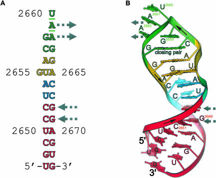
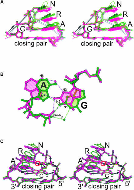
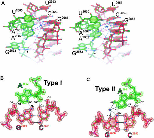
GNRA tetraloops (N is A, C, G, or U; R is A or G) are basic building blocks of RNA structure that often interact with proteins or other RNA structural elements. Understanding sequence-dependent structural variation among different GNRA tetraloops is an important step toward elucidating the molecular basis of specific GNRA tetraloop recognition by proteins and RNAs. Details of the geometry and hydration of this motif have been based on high-resolution crystallographic structures of the GRRA subset of tetraloops; less is known about the GYRA subset (Y is C or U). We report here the structure of a GUAA tetraloop determined to 1.4 A resolution to better define these details and any distinctive features of GYRA tetraloops. The tetraloop is part of a 27-nt structure that mimics the universal sarcin/ricin loop from Escherichia coli 23S ribosomal RNA in which a GUAA tetraloop replaces the conserved GAGA tetraloop. The adenosines of the GUAA tetraloop form an intermolecular contact that is a commonplace RNA tertiary interaction called an A-minor motif. This is the first structure to reveal in great detail the geometry and hydration of a GUAA tetraloop and an A-minor motif. Comparison of tetraloop structures shows a common backbone geometry for each of the eight possible tetraloop sequences and suggests a common hydration. After backbone atom superposition, equivalent bases from different tetraloops unexpectedly depart from coplanarity by as much as 48 degrees. This variation displaces the functional groups of tetraloops implicated in protein and RNA binding, providing a recognition feature.
Figures
Similar articles
-
Comprehensive features of natural and in vitro selected GNRA tetraloop-binding receptors.Nucleic Acids Res. 2008 Mar;36(4):1138-52. doi: 10.1093/nar/gkm1048. Epub 2007 Dec 23. Nucleic Acids Res. 2008. PMID: 18158305 Free PMC article.
-
UNAC tetraloops: to what extent do they mimic GNRA tetraloops?Biopolymers. 2012 Aug;97(8):617-28. doi: 10.1002/bip.22049. Biopolymers. 2012. PMID: 22605553
-
Incorporating a Thiophosphate Modification into a Common RNA Tetraloop Motif Causes an Unanticipated Stability Boost.Biochemistry. 2020 Dec 15;59(49):4627-4637. doi: 10.1021/acs.biochem.0c00685. Epub 2020 Dec 4. Biochemistry. 2020. PMID: 33275419
-
Recognition modes of RNA tetraloops and tetraloop-like motifs by RNA-binding proteins.Wiley Interdiscip Rev RNA. 2014 Jan-Feb;5(1):49-67. doi: 10.1002/wrna.1196. Epub 2013 Oct 3. Wiley Interdiscip Rev RNA. 2014. PMID: 24124096 Free PMC article. Review.
-
An RNA folding motif: GNRA tetraloop-receptor interactions.Q Rev Biophys. 2013 Aug;46(3):223-64. doi: 10.1017/S0033583513000048. Epub 2013 Aug 5. Q Rev Biophys. 2013. PMID: 23915736 Review.
Cited by
-
Identification and characterization of RNA pentaloop sequence families.NAR Genom Bioinform. 2023 Jan 10;5(1):lqac102. doi: 10.1093/nargab/lqac102. eCollection 2023 Mar. NAR Genom Bioinform. 2023. PMID: 36632613 Free PMC article.
-
The GANC tetraloop: a novel motif in the group IIC intron structure.J Mol Biol. 2008 Nov 14;383(3):475-81. doi: 10.1016/j.jmb.2008.08.043. Epub 2008 Aug 26. J Mol Biol. 2008. PMID: 18773908 Free PMC article.
-
Comprehensive features of natural and in vitro selected GNRA tetraloop-binding receptors.Nucleic Acids Res. 2008 Mar;36(4):1138-52. doi: 10.1093/nar/gkm1048. Epub 2007 Dec 23. Nucleic Acids Res. 2008. PMID: 18158305 Free PMC article.
-
Structural Basis of Detection and Signaling of DNA Single-Strand Breaks by Human PARP-1.Mol Cell. 2015 Dec 3;60(5):742-754. doi: 10.1016/j.molcel.2015.10.032. Epub 2015 Nov 25. Mol Cell. 2015. PMID: 26626479 Free PMC article.
-
Structural features of a 3' splice site in influenza a.Biochemistry. 2015 Jun 2;54(21):3269-85. doi: 10.1021/acs.biochem.5b00012. Epub 2015 May 21. Biochemistry. 2015. PMID: 25909229 Free PMC article.
References
-
- Abramovitz, D.L., Friedman, R.A., and Pyle, A.M. 1996. Catalytic role of 2′-hydroxyl groups within a group II intron active site. Science 271: 1410–1413. - PubMed
-
- Ban, N., Nissen, P., Hansen, J., Moore, P.B., and Steitz, T.A. 2000. The complete atomic structure of the large ribosomal subunit at 2.4 Å resolution. Science 289: 905–920. - PubMed
-
- Batey, R.T., Rambo, R.P., Lucast, L., Rha, B., and Doudna, J.A. 2000. Crystal structure of the ribonucleoprotein core of the signal recognition particle. Science 287: 1232–1239. - PubMed
-
- Batey, R.T., Sagar, M.B., and Doudna, J.A. 2001. Structural and energetic analysis of RNA recognition by a universally conserved protein from the signal recognition particle. J. Mol. Biol. 307: 229–246. - PubMed
-
- Brunger, A.T. 1992. X-PLOR Version 3.1: A system for X-ray crystallography and NMR. Yale University Press, New Haven, CT.
Publication types
MeSH terms
Substances
Grants and funding
LinkOut - more resources
Full Text Sources
Molecular Biology Databases
Research Materials