Comparative Study
. 2003 Mar;12(3):426-37.
doi: 10.1110/ps.0233503.

X-ray structure of the AAC(6')-Ii antibiotic resistance enzyme at 1.8 A resolution; examination of oligomeric arrangements in GNAT superfamily members

Affiliations
Comparative Study

X-ray structure of the AAC(6')-Ii antibiotic resistance enzyme at 1.8 A resolution; examination of oligomeric arrangements in GNAT superfamily members

David L Burk et al. Protein Sci. 2003 Mar.

Abstract

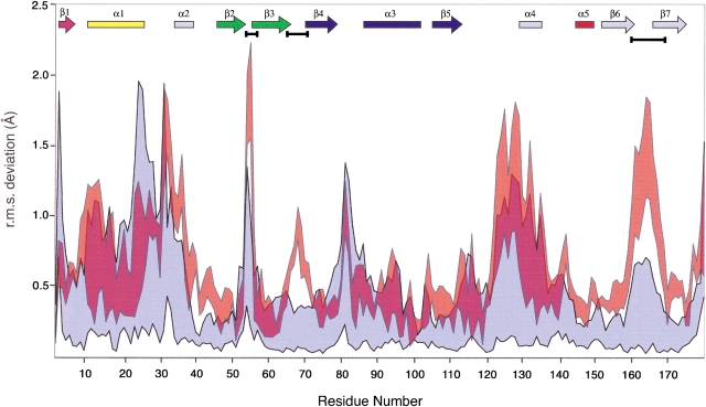
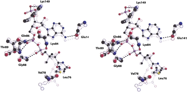
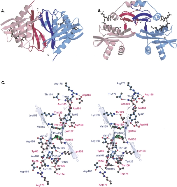
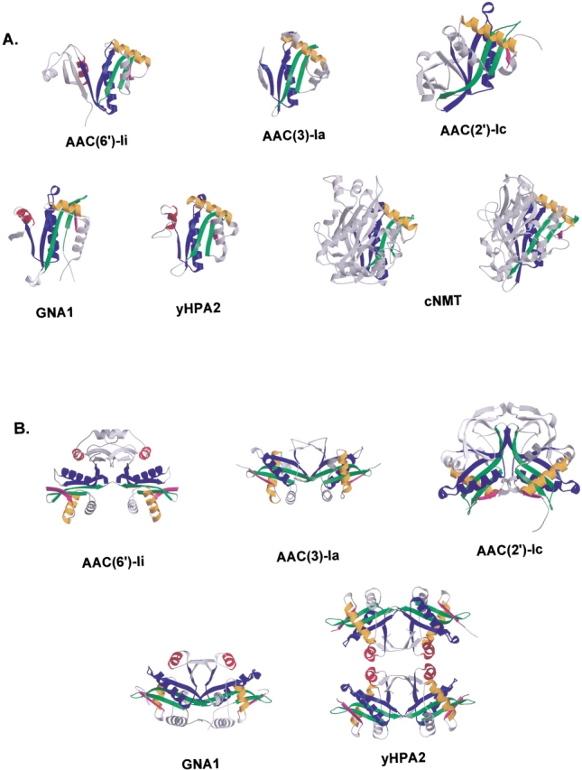
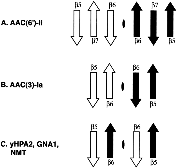
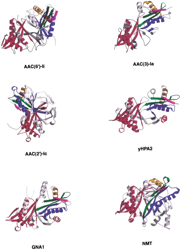
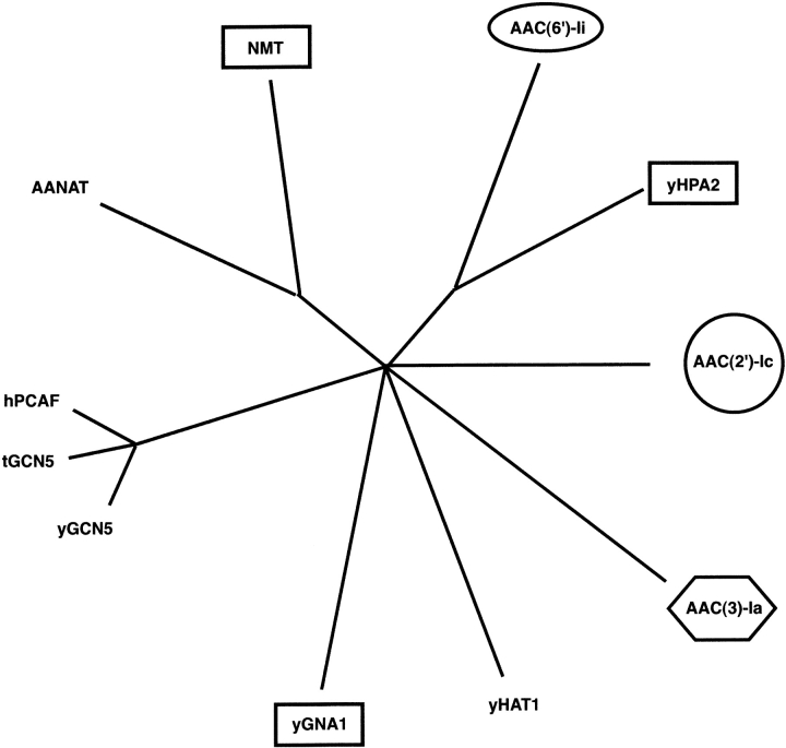
The rise of antibiotic resistance as a public health concern has led to increased interest in studying the ways in which bacteria avoid the effects of antibiotics. Enzymatic inactivation by several families of enzymes has been observed to be the predominant mechanism of resistance to aminoglycoside antibiotics such as kanamycin and gentamicin. Despite the importance of acetyltransferases in bacterial resistance to aminoglycoside antibiotics, relatively little is known about their structure and mechanism. Here we report the three-dimensional atomic structure of the aminoglycoside acetyltransferase AAC(6')-Ii in complex with coenzyme A (CoA). This structure unambiguously identifies the physiologically relevant AAC(6')-Ii dimer species, and reveals that the enzyme structure is similar in the AcCoA and CoA bound forms. AAC(6')-Ii is a member of the GCN5-related N-acetyltransferase (GNAT) superfamily of acetyltransferases, a diverse group of enzymes that possess a conserved structural motif, despite low sequence homology. AAC(6')-Ii is also a member of a subset of enzymes in the GNAT superfamily that form multimeric complexes. The dimer arrangements within the multimeric GNAT superfamily members are compared, revealing that AAC(6')-Ii forms a dimer assembly that is different from that observed in the other multimeric GNAT superfamily members. This different assembly may provide insight into the evolutionary processes governing dimer formation.


Figures

Figure 1.
Figure 1.
This graph illustrates the maximum and minimum values of the r.m.s.d in main-chain atomic position following superposition of the four copies of the AAC(6′)-Ii•CoA monomer present in the unit cell (blue). Also plotted on the same axes is the corresponding range of RMSD values for the superposition of the AAC(6′)-Ii•CoA and AAC(6′)-Ii•AcCoA structures (red). The secondary structural elements are indicated at the top of the frame (colored according to Wybenga-Groot et al. 1999). Gaps between the colored regions in this plot indicate areas of the polypeptide chain in which the deviation in atomic position between the CoA and AcCoA structures exceeds that observed between the four copies of the CoA monomer in the unit cell. These regions are highlighted with horizontal black bars below the secondary structural elements.
Figure 2.
Figure 2.
A stereo ball-and-stick representation of the substrate binding site of the AAC(6′)-Ii enzyme. The figure includes both the complex with CoA (full color) and the superimposed structure of the AcCoA complex (partially transparent). The figure includes those residues that form hydrogen bond interactions with the cofactor (represented by broken lines). Carbon atoms are depicted in black, oxygen atoms in red, nitrogen atoms in blue, and sulfur atoms in yellow.
Figure 3.
Figure 3.
(A) A cartoon figure showing two views of the AAC(6′)-Ii•CoA dimer. One monomer is colored blue and the other red, with the secondary structural elements involved in the dimer interface represented by a darker shade of their respective colors. (B) This view of the AAC(6′) dimer obtained by rotating the dimer in Figure 3A ▶ by 90° about a horizontal axis passing through the center of the dimer. Elements that are in the background in (A) are now at the top of (B). The cofactors are shown as black ball-and-stick models. (C) A ball-and-stick figure depicting the interactions between the monomers of the AAC(6′) dimer. The color scheme of the panel is similar to that of (A) and (B), with the amino acid residues from one monomer colored red and those from the other colored blue. Hydrogen bonds are shown as dashed lines. Residue Phe130, a aromatic residue involved in an interdimer stacking interaction is shown in green. The partially transparent backbone atoms and cartoon β-strands indicate the position and direction of the β6 strands of the two AAC(6′)-Ii monomers.
Figure 4.
Figure 4.
(A) The first panel of this figure shows cartoon representations of the monomer forms of the GNAT superfamily members that are known to be multimeric. In each case, the conserved structural features are colored according to Wybenga-Groot et al. (1999), while the remaining parts of the molecules are represented in gray. All structures have been superimposed such that the conserved motifs are in similar orientations. Although not multimeric, NMT is included here because it contains two copies of the GNAT conserved motif. (B) The second panel shows cartoon representations of the multimeric GNAT superfamily members. Figures are colored as in (A) and are oriented such that the two copies of the GNAT motif and the C-terminal sections of the assemblies can be clearly seen.
Figure 5.
Figure 5.
Schematic figure illustrating the topology of the β-strands involved in the central interdimer interactions in the multimeric members of the GNAT superfamily of enzymes. Strands from one monomer are white, while those from the second are shaded black. The dimer interface is represented by a black oval.
Figure 6.
Figure 6.
Cartoon representations of the four multimeric GNAT superfamily members and the NMT monomer containing two GNAT motifs. In each, one copy of the motif has been colored red, and the other according to the scheme of Wybenga-Groot et al. (1999). The remainder of the protein is shown in gray. Each molecule has been oriented such that the red motifs are superimposed, illustrating the different relative orientations of the conserved GNAT motifs among the superfamily members.
Figure 7.
Figure 7.
An unrooted quartet puzzling tree showing the most probable evolutionary relationship between the 10 GNAT superfamily members whose three-dimensional structures are known. The tree is based on a structure-based amino acid sequence alignment prepared by superimposing the atomic coordinates of the proteins with those of AAC(6)-Ii. Of the residues identified as structurally similar, only those residues that were common to all 10 proteins were used in the subsequent phylogenetic analysis. Oligomeric proteins are indicated by boxes, ovals, hexagons, and circles with identical shapes indicating proteins with similar dimer arrangements. The maximum likelihood program Tree-Puzzle was used, with default settings and AAC(2′)-Ic as outgroup, to produce the evolutionary tree (Strimmer and von Haeseler 1996). The result was then visualized with the program TreeView (Page 1996). The length of the lines is proportional to the maximum liklihood branch length and reflects the degree of sequence difference.

References

    1. Angus-Hill, M.L., Dutnall, R.N., Tafrov, S.T., Sternglanz, R., and Ramakrishnan, V. 1999. Crystal structure of the histone acetyltransferase Hpa2: A tetrameric member of the Gcn5-related N-acetyltransferase superfamily. J. Mol. Biol. 294 1311–1325. - PubMed
    1. Bennett, M.J., Schlunegger, M.P., and Eisenberg, D. 1995. 3D domain swapping: A mechanism for oligomer assembly. Protein Sci. 5 2455–2468. - PMC - PubMed
    1. Berman, H.M., Westbrook, J., Feng, Z., Gilliland, G., Bhat, T.N., Weissig, H., Shindyalov, I.N., and Bourne, P.N. 2000. The Protein Data Bank. Nucleic Acids Res. 28 235–242. - PMC - PubMed
    1. Bhatnagar, R.S., Futterer, K., Farazi, T.A., Korolev, S., Murray, C.L., Jackson-Machelski, E., Gokel, G.W., Gordon, J.I., and Waksman, G. 1998. Structure of N-myrisoyltransferase with bound myristoylCoA and peptide substrate analogs. Nat. Struct. Biol. 5 1091–1097. - PubMed
    1. Brünger, A., Adams, P.D., Clore, G.M., DeLano, W.L., Gros, P., GrosseKunstleve, R.W., Jiang, J-S., Kuszewski, J., Nilges, M., Pannu, N.S., et al. 1998. Crystallography & NMR system: A new software suite for macromolecular structure determination. Acta Crystallogr. D54 905–921. - PubMed

Publication types