. 2003 Mar;72(3):681-90.
doi: 10.1086/368294.

Mutations in genes encoding fast-twitch contractile proteins cause distal arthrogryposis syndromes

Affiliations

Mutations in genes encoding fast-twitch contractile proteins cause distal arthrogryposis syndromes

Sandy S Sung et al. Am J Hum Genet. 2003 Mar.

Abstract

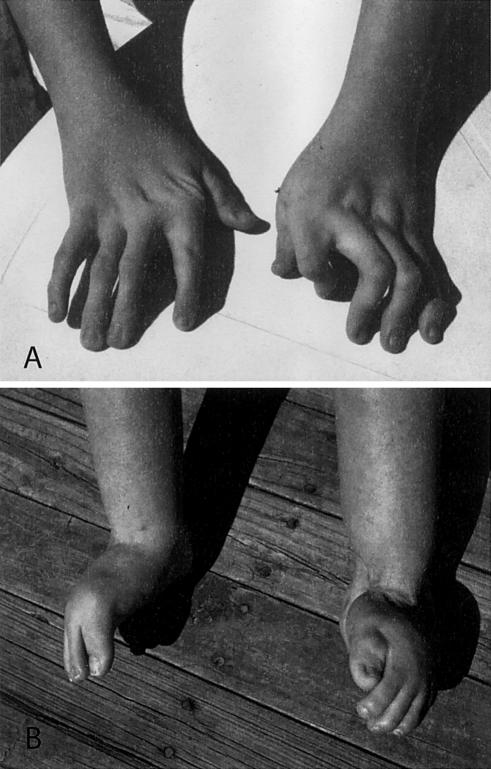
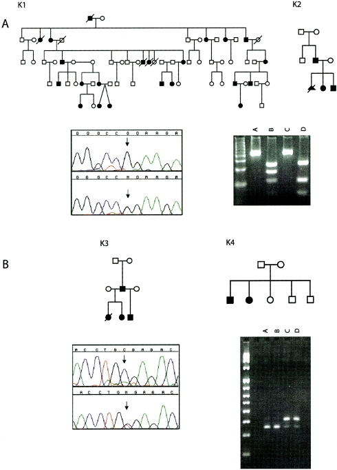
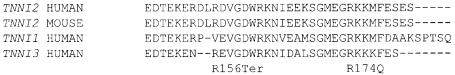
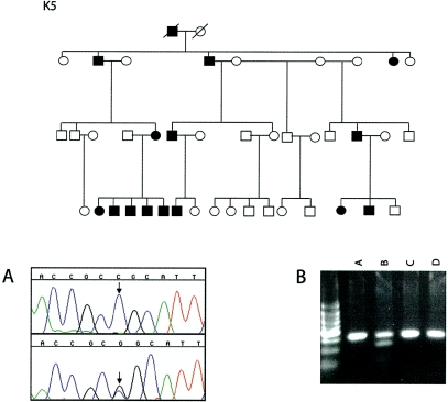
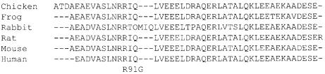
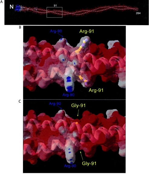
The distal arthrogryposes (DAs) are a group of disorders characterized by multiple congenital contractures of the limbs. We previously mapped a locus for DA type 2B (DA2B), the most common of the DAs, to chromosome 11. We now report that DA2B is caused by mutations in TNNI2 that are predicted to disrupt the carboxy-terminal domain of an isoform of troponin I (TnI) specific to the troponin-tropomyosin (Tc-Tm) complex of fast-twitch myofibers. Because the DAs are genetically heterogeneous, we sought additional candidate genes by examining modifiers of mutant Drosophila isoforms of TnI. One of these modifiers, Tm2, encodes tropomyosin, another component of the Tc-Tm complex. A human homologue of Tm2, TPM2, encodes beta-tropomyosin and maps to the critical interval of DA type 1 (DA1). We discovered that DA1 is caused by substitution of a highly conserved amino acid residue in beta-tropomyosin. These findings suggest that DAs, in general, may be caused by mutations in genes encoding proteins of the contractile apparatus specific to fast-twitch myofibers. This provides a new opportunity to directly study the etiology and pathogenesis of multiple-congenital-contracture syndromes.


Figures

Figure  1
Figure 1
Typical malformations observed in individuals with DA2B. A, Hands characterized by camptodactyly and ulnar deviation. B, Feet showing camptodactyly accompanied by calcaneovalgus deformities and a vertical talus or clubfoot (not shown).
Figure  2
Figure 2
Electropherograms of mutations in exon 8 of TNNI2. A, G→A missense mutation at position 521 in exon 8 of TNNI2 in kindreds K1 and K2. The mutation creates a novel MspI restriction site. Digests of TNNI2 amplicons from an affected individual (lane B) fractionate into four fragments (243 bp, 166 bp, 110 bp, and 56 bp), whereas only three fragments (243 bp, 110 bp, and 56 bp) are observed in an unaffected individual (lane D). Lanes A and C, Undigested control samples. B, C→T nonsense mutation at bp 466 of exon 8 of TNNI2 in kindreds K3 and K4. The mutation eliminates a BfuAI restriction site. Digests of TNNI2 amplicons from two affected sibs in kindred K4 (lanes C and D) fractionate into two fragments (409 bp and 344 bp), whereas only one fragment (344 bp) is found in the unaffected parents (lanes A and B). The 65-bp product cannot be observed on this gel.
Figure  3
Figure 3
Alignment of genes encoding isoforms of TnI specific to fast-twitch muscle fibers (TNNI2) in humans and mice, slow-twitch muscle fibers (TNNI1), and cardiac muscle (TNNI3). The positions of the R156Ter and R174Q mutations are indicated.
Figure  4
Figure 4
Top, Pedigree of family K5. Bottom, electropherogram of a mutation on exon 3 and digests of amplicons. A, Electropherogram of a C→G missense mutation at position 271 in exon 3 of TPM2. The mutation creates a novel SacII restriction site in kindred K5. B, Digests of TPM2 amplicons from an affected individual. The digests from this individual separate into three fragments (320 bp, 218 bp, and 102 bp) (lane B), whereas amplicons from an unaffected individual remain undigested (lane D). Lanes A and C, Undigested control amplicons.
Figure  5
Figure 5
Alignment of isoforms of β-tropomyosin from six animal species. The R91G substitution replaces an arginine residue that is invariant among species.
Figure  6
Figure 6
A, Homology model of human β-tropomyosin dimer with a molecular surface colored by relative surface electrostatic charge. The model is based on the 7-Å crystal structure of porcine cardiac α-tropomyosin dimer (Protein Database entry 1C1G). A partially transparent molecular surface is shown colored by electrostatic charge: red indicates regions dominated by negative charge; white indicates areas that are neutral; blue indicates positive charge. Relatively few positively charged arginine and lysine surface amino acid side chains are found outside the N-terminal region. The sparse positively charged amino acids are largely neutralized by the large number of surface negatively charged aspartate and glutamate amino acids, which creates the dominant negatively charged surface character of the molecule. A ribbon model (gray) shows the characteristic coiled-coil fold of tropomyosin and is visible through the partially transparent molecular surface. “N” indicates the relatively positively charged N-terminal region, which is important for end-to-end polymerization, and “284” indicates the C-terminus. Box indicates the region of the molecule surrounding residue 91 shown in detail (panels B and C). B, Detailed view of the area of surrounding residue 91. A ball-and-stick model of the Arg91 side chain is colored by atom type and is visible through the partially transparent molecular surface. The positive charge provided by Arg90 and Arg91 is flanked by negative charge, characteristic of the molecule as a whole. C, Detailed view of the area surrounding residue 91 with R91G mutation. Mutation to glycine neutralizes the positive charge at that position, leading to an increase in local negatively charged character, seen as an increase in red color in panel C relative to panel B. Figures were generated with the program Swiss PDB Viewer.

References

Electronic-Database Information

    1. Online Mendelian Inheritance in Man (OMIM) http://www.ncbi.nlm.nih.gov/Omim/ (for DA1 [MIM 108120], DA2B [MIM 601680], and FSS [MIM 193700])

References

    1. Andrade MD, Barnholtz JS, Amos CI, Lockmiller C, Scott A, Risman M, Hecht J (1998) Segregation analysis of idiopathic talipes equinovarus in a Texan population. Am J Med Genet 79:97–102 - PubMed
    1. Bamshad M, Bohnsack JF, Jorde LB, Carey JC (1996a) Distal arthrogryposis type 1: clinical analysis of a large kindred. Am J Med Genet 65:282–285 - PubMed
    1. Bamshad M, Jorde LB, Carey JC (1996b) A revised and extended classification of the distal arthrogryposes. Am J Med Genet 65:277–281 - PubMed
    1. Bamshad M, Watkins WS, Zenger RK, Bohnsack JF, Carey JC, Otterud B, Krakowiak PA, Robertson M, Jorde LB (1994) A gene for distal arthrogryposis type 1 maps to the pericentromeric region of chromosome 9. Am J Hum Genet 55:1153–1158 - PMC - PubMed
    1. Beall CJ, Fyrberg E (1991) Muscle abnormalities in Drosophila melanogaster heldup mutants are caused by missing or aberrant troponin-I isoforms. J Cell Biol 114:941–951 - PMC - PubMed

Publication types

MeSH terms

Associated data