

Retinal findings in systemic sclerosis: a comparison with nailfold capillaroscopic patterns
- PMID: 12594103
- PMCID: PMC1754455
- DOI: 10.1136/ard.62.3.204
Retinal findings in systemic sclerosis: a comparison with nailfold capillaroscopic patterns
Abstract
Objective: To determine the prevalence of retinal disease in systemic sclerosis (SSc) and to characterise the findings of retinopathy. Additionally, to analyse the association of retinal disease with other clinical/laboratory findings, particularly the findings of nailfold capillaries in patients with SSc.
Methods: Photographs of the ocular fundi were taken and were evaluated by an ophthalmologist who was unaware of the SSc status of the patients. The nailfold capillaries were analysed with a dermatoscope. Patients were divided into two groups according to the presence (group A) or absence (group B) of retinal disease.
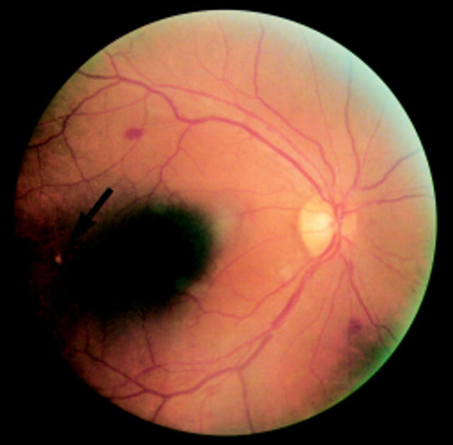
Results: Retinal findings of the patients with SSc consisted of hard exudates, vascular tortuosity, microhaemorrhage, and macular degeneration. The prevalence of retinal disease among the patients with SSc was 34% (10/29), compared with 8%(3/38) among the controls (p=0.011). The mean systolic blood pressure and the age of the patients in group A were significantly higher than those in group B. However, there was no significant difference in the nailfold capillary damage between groups A and B.
Conclusion: Retinal abnormalities are often seen in patients with SSc and they may reflect the vascular changes characteristic of SSc. However, retinal changes may differ in quality from the changes of nailfold capillaries.
Figures
Similar articles
-
Nailfold capillaroscopic changes in patients with idiopathic pulmonary arterial hypertension and systemic sclerosis-related pulmonary arterial hypertension.Microvasc Res. 2017 Nov;114:46-51. doi: 10.1016/j.mvr.2017.06.005. Epub 2017 Jun 12. Microvasc Res. 2017. PMID: 28619664
-
Nailfold capillaroscopic findings in systemic sclerosis related lung fibrosis and in idiopathic lung fibrosis.Joint Bone Spine. 2010 Dec;77(6):570-4. doi: 10.1016/j.jbspin.2010.02.019. Epub 2010 May 23. Joint Bone Spine. 2010. PMID: 20547086
-
Microvascular involvement in systemic sclerosis: capillaroscopic findings.Semin Arthritis Rheum. 2001 Jun;30(6):397-402. doi: 10.1053/sarh.2001.20269. Semin Arthritis Rheum. 2001. PMID: 11404822
-
Microvascular damage evaluation in systemic sclerosis: the role of nailfold videocapillaroscopy and laser techniques.Reumatismo. 2017 Dec 21;69(4):147-155. doi: 10.4081/reumatismo.2017.959. Reumatismo. 2017. PMID: 29320840 Review.
-
The emerging application of semi-quantitative and quantitative capillaroscopy in systemic sclerosis.Microvasc Res. 2018 Jul;118:113-120. doi: 10.1016/j.mvr.2018.03.004. Epub 2018 Mar 12. Microvasc Res. 2018. PMID: 29544760 Review.
Cited by
-
Acute retinal artery occlusion in systemic sclerosis: a rare manifestation of systemic sclerosis fibroproliferative vasculopathy.Semin Arthritis Rheum. 2013 Oct;43(2):204-8. doi: 10.1016/j.semarthrit.2012.12.025. Epub 2013 Feb 19. Semin Arthritis Rheum. 2013. PMID: 23433487 Free PMC article.
-
Prevalence of ocular manifestations in systemic sclerosis patients.Arch Med Sci. 2013 Dec 30;9(6):1107-13. doi: 10.5114/aoms.2013.39217. Epub 2013 Nov 29. Arch Med Sci. 2013. PMID: 24482658 Free PMC article.
-
Vascular Endothelial Damage in COPD: Where Are We Now, Where Will We Go?Diagnostics (Basel). 2024 Apr 30;14(9):950. doi: 10.3390/diagnostics14090950. Diagnostics (Basel). 2024. PMID: 38732364 Free PMC article. Review.
-
Retinal microvasculature alteration in patients with systemic sclerosis and chloroquine treatment.Quant Imaging Med Surg. 2022 Oct;12(10):4885-4899. doi: 10.21037/qims-21-1166. Quant Imaging Med Surg. 2022. PMID: 36185048 Free PMC article.
-
A multimodal ophthalmic analysis in patients with systemic sclerosis using ocular response analyzer, corneal topography and specular microscopy.Int Ophthalmol. 2020 Feb;40(2):287-296. doi: 10.1007/s10792-019-01173-x. Epub 2019 Sep 28. Int Ophthalmol. 2020. PMID: 31564047
References
MeSH terms
LinkOut - more resources
Full Text Sources
Medical
