. 2003 Mar 4;100(5):2718-23.
doi: 10.1073/pnas.0437841100. Epub 2003 Feb 19.

RNA interference of influenza virus production by directly targeting mRNA for degradation and indirectly inhibiting all viral RNA transcription

Affiliations

RNA interference of influenza virus production by directly targeting mRNA for degradation and indirectly inhibiting all viral RNA transcription

Qing Ge et al. Proc Natl Acad Sci U S A. .

Abstract

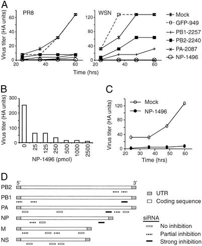
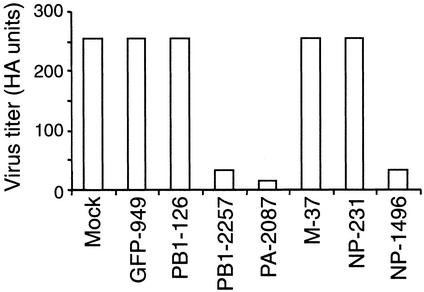
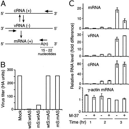
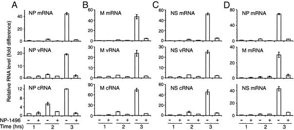
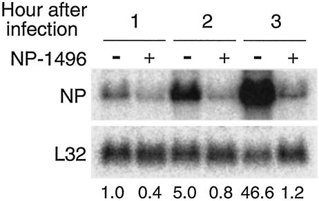
Influenza A virus causes widespread infection in the human respiratory tract, but existing vaccines and drug therapy are of limited value. Here we show that short interfering RNAs (siRNAs) specific for conserved regions of the viral genome can potently inhibit influenza virus production in both cell lines and embryonated chicken eggs. The inhibition depends on the presence of a functional antisense strand in the siRNA duplex, suggesting that viral mRNA is the target of RNA interference. However, siRNA specific for nucleocapsid (NP) or a component of the RNA transcriptase (PA) abolished the accumulation of not only the corresponding mRNA but also virion RNA and its complementary RNA. These siRNAs also broadly inhibited the accumulation of other viral, but not cellular, RNAs. The findings reveal that newly synthesized NP and PA proteins are required for influenza virus transcription and replication and provide a basis for the development of siRNAs as prophylaxis and therapy for influenza infection in humans.


Figures

Figure 1
Figure 1
siRNAs interfere with influenza A virus production in MDCK cells. (A) Inhibition of influenza virus production in MDCK cells by selected siRNAs. MDCK cells were first electroporated with siRNAs and then infected 8 h later with PR8 or WSN virus at a moi of 0.01. Viral titers in the culture supernatants at different times after infection were measured by HA assay. HA units are arithmetic means based on titer endpoints of arithmetic dilutions. Virus titers (HA units) from five siRNA-treated cultures are shown over time for both PR8 (Left) and WSN (Right) infections. NP-1496, etc., are siRNAs specific for different viral genes. For example, NP-1496 indicates an siRNA specific for NP, with the beginning nucleotide at position 1496 of the NP sequence. (B) Inhibition of influenza virus production by different doses of siRNA. MDCK cells were transfected with the indicated amount of NP-1496 followed by infection 8 h later with PR8 virus at a moi of 0.1. Virus titer was measured 48 h after infection. Data shown are from one of two experiments. (C) Inhibition of influenza virus production by siRNA administered after virus infection. MDCK cells were infected with PR8 virus at a moi of 0.01 and were electroporated 2 h later with NP-1496 (2.5 nmol). Virus titer was measured at the indicated time after infection. Data shown are from one of two experiments. (D) Schematic diagrams showing the location of each siRNA in the viral genome and its relative potency in inhibiting influenza virus production in MDCK cells (based on data in Table 1). UTR, untranslated regions.
Figure 2
Figure 2
siRNAs interfere with influenza virus production in embryonated chicken eggs. A mixture of siRNAs (2.5 nmol), Oligofectamine, and PR8 virus (500 pfu) was injected into the allantoic cavity of 10-day-old embryonated chicken eggs. Allantoic fluid was collected 17 h later and assayed for virus titers as in Fig. 1. Data shown are from one of two experiments.
Figure 3
Figure 3
mRNA is the likely target for siRNA-mediated interference. (A) Schematic diagram illustrating the relationship among influenza virus vRNA, mRNA, and cRNA. Whereas cRNA is the exact complement of vRNA, mRNA contains a cap structure at the 5′ end (not shown) and a poly(A) sequence that occurs at a site 15–22 nt before the 5′ end of the vRNA segment. Arrows indicate the positions of primers used to distinguish among the various RNAs during RT. (B) Inhibition of influenza virus production requires a wild-type (wt) antisense strand in the duplex siRNA. MDCK cells were first electroporated with siRNAs formed from wt and modified (m) strands and were infected 8 h later with PR8 virus at a moi of 0.1. Virus titers in the culture supernatants were assayed 24 h after infection as in Fig. 1. Data shown are from one of two experiments. (C) M-specific siRNA inhibits the accumulation of specific mRNA. MDCK cells were electroporated with M-37, infected with PR8 virus at a moi of 0.01, and harvested for RNA isolation 1, 2, and 3 h after infection. The levels of M-specific mRNA, cRNA, and vRNA were measured by RT using RNA-specific primers as indicated in A, followed by real-time PCR. The level of each viral RNA species was normalized to the level of γ-actin mRNA (Bottom) in the same sample. The relative levels of RNAs are shown as mean value ± SD. Data shown are from one of two experiments.
Figure 4
Figure 4
NP-specific siRNA inhibits the accumulation of not only NP- but also M- and NS-specific mRNA, vRNA, and cRNA. MDCK (A–C) and Vero (D) cells were electroporated with NP-1496, infected with PR8 virus at a moi of 0.1, and harvested for RNA isolation 1, 2, and 3 h after infection. The levels of mRNA, cRNA, and vRNA specific for NP, M, and NS were measured by RT using RNA-specific primers, followed by real-time PCR. The level of each viral RNA species is normalized to the level of γ-actin mRNA (data not shown) in the same sample. The relative levels of RNAs are shown. Data shown are from one of two experiments.
Figure 5
Figure 5
NP-specific siRNA inhibits the accumulation of all NP viral RNAs. MDCK cells were electroporated with NP-1496, infected with PR8 virus at a moi of 0.1, and harvested for RNA isolation 1, 2, and 3 h after infection. Total RNA (15 μg) was separated by a 1.2% denaturing agarose gel, transferred, and hybridized sequentially with probes specific for NP (Upper) and the ribosomal L32 gene (Lower). The numbers indicate the relative levels of NP-specific RNA normalized to the levels of L32. The level of NP-specific RNA in cells that were infected for 1 h in the absence of siRNA is arbitrarily given a value of 1. The NP-specific probe was double-stranded and hybridized to mRNA, vRNA, and cRNA.

References

    1. Lamb R A, Krug R M. In: Fundamental Virology. Knipe D M, Howley P M, editors. Philadelphia: Lippincott Williams & Wilkins; 2001. pp. 725–770.
    1. Fukuda K, Bridges C B, Brammer T L, Izurieta H S, Cox N J. Morbid Mortal Wkly Rep. 1999;48:1–28.
    1. Patterson K D, Pyle G F. Bull Hist Med. 1991;65:4–21. - PubMed
    1. Parvin J D, Moscona A, Pan W T, Leider J M, Palese P. J Virol. 1986;59:377–383. - PMC - PubMed
    1. Webster R G, Laver W G, Air G M, Schild G C. Nature. 1982;296:115–121. - PubMed

Publication types

MeSH terms