. 2003 Feb;52(2):89-96.
doi: 10.1007/s00262-002-0362-7. Epub 2003 Jan 21.

The abl/bcr gene product as a novel leukemia-specific antigen: peptides spanning the fusion region of abl/bcr can be recognized by both CD4+ and CD8+ T lymphocytes

Affiliations

The abl/bcr gene product as a novel leukemia-specific antigen: peptides spanning the fusion region of abl/bcr can be recognized by both CD4+ and CD8+ T lymphocytes

Wolfgang M Wagner et al. Cancer Immunol Immunother. 2003 Feb.

Abstract

Chronic myelogenous leukemia (CML) is characterized by a reciprocal translocation leading to the Philadelphia chromosome. Two fusion genes are created by this translocation: bcr/abl and abl/bcr. The fusion regions of both translocation products are unique and strictly limited to leukemia cells, giving rise to potential tumor-specific antigens. Although several studies on the immunogenicity of peptides spanning the bcr/abl fusion region have been reported, little is known about the corresponding reciprocal translocation product abl/bcr. Here we report that synthetic peptides representing the fusion region of the abl/bcr forms a1bb3 and a1bb4 can be specifically recognized by HLA-A2-restricted cytotoxic T lymphocytes from healthy donors. Furthermore, HLA-matched a1bb3-expressing CML cells can be recognized by a1bb3-specific HLA-A2-restricted T cells, indicating natural processing and presentation of abl/bcr protein by leukemia cells. Moreover, a 19-mer peptide encompassing this class I-binding sequence also elicited a1bb3-specific class II-restricted T-cell responses. Thus, both class I- and class II-restricted T-cell responses can be stimulated in healthy donors by abl/bcr peptides in vitro. Because abl/bcr is expressed in the majority of CML patients, it may represent a highly leukemia-specific antigen with potential use in immunotherapy.


Figures

Fig. 1a, b.
Fig. 1a, b.
Expression of abl/bcr and bcr/abl mRNA in PBMC of 12 CML patients. a abl/bcr. Length of fragments: a1bb3 342 bp, a1bb4 267 bp. b bcr/abl. Length of fragments: b2a2 404 bp, b3a2 479 bp (M 100-bp ladder, lanes 1–12 CML1–CML12, B BV-173, E EM-2, K K-562, H H2Odd)
Fig. 2a, b.
Fig. 2a, b.
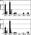
Recognition of CML cells by a1bb3 peptide-specific T cells is dependent on HLA-A2 and CD8 expression. TNFα release after coincubation of a1bb3 peptide-specific T cell lines B360 and B363 with HLA-A*0201-positive CML cells (black bars) could be blocked with monoclonal antibody BB7.2 against HLA-A2 (hatched bars) or OKT8 against CD8 (stippled bars). Spontaneous release of TNFα by T cells was below the level of detection, and spontaneous release by CML cells is shown in the white bars (a B360, b B363). (CML 9, 11 HLA-A*0201+/a1bb3 transcript+; CML 26 HLA-A*0201/a1bb3 transcript+; CML 8, 24, 25 HLA-A*0201+/a1bb4 transcript+)
Fig. 3a, b.
Fig. 3a, b.
a1bb4 peptide-specific T cells fail to recognize HLA-matched a1bb4-expressing CML cells. HLA-A*0201-restricted T cell lines B460 and B463, specific for the a1bb4 peptide, were coincubated with various CML cells (black bars). After 24 h the TNF-α content in the supernatant was determined by ELISA. No significant T cell response was observed either to a1bb4-expressing or to a1bb3-expressing CML cells. The addition of blocking antibodies against HLA-A2 (hatched bars) or CD8 (stippled bars) had no effect on TNF-α release. Spontaneous release of CML cells is shown in the white bars (a B460, b B463). (CML 9, 11 HLA-A*0201+/a1bb3 transcript+; CML 26 HLA-A*0201/a1bb3 transcript+; CML 8, 24, 25 HLA-A*0201+/a1bb4 transcript+)
Fig. 4.
Fig. 4.
CD4+ T cell clones specifically recognize autologous PBMC pulsed with an a1bb3 peptide. T cells from a healthy donor sensitized in vitro against a 19mer a1bb3 peptide were cloned by limiting dilution. T cell clones respond specifically to autologous PBMC pulsed with the a1bb3 peptide (black bars), but not to PBMC alone (white bars) or to PBMC pulsed with a control peptide (hatched bars)
Fig. 5a, b.
Fig. 5a, b.
Recognition of a1bb3 peptide by CD4+ T cells is HLA-DR-restricted and dose-dependent. a Autocrine proliferation of TCC 9-8-3 in response to autologous PBMC pulsed with increasing amounts of 19mer a1bb3 (■) or irrelevant control peptide (□). b Autocrine proliferation of HLA-DRβ1*0701 homozygous TCC 9-8-3 in response to autologous PBMC pulsed with 19mer a1bb3 is blocked by preincubating stimulator cells with an anti-HLA-DR antibody (L243), but not with antibodies against HLA-DQ (TÜ22) or HLA-DP (B7/21)

Similar articles

Cited by

References

    1. Goldman Blood. 2001;98:2039. doi: 10.1182/blood.V98.7.2039. - DOI - PubMed
    1. Gorre Curr Opin Hematol. 2002;9:303. doi: 10.1097/00062752-200207000-00007. - DOI - PubMed
    1. Smit Proc Natl Acad Sci U S A. 1998;95:10152. doi: 10.1073/pnas.95.17.10152. - DOI - PMC - PubMed
    1. Deininger Blood. 2000;96:3343. - PubMed
    1. Faderl N Engl J Med. 1999;341:164. - PubMed

Publication types

MeSH terms