. 2003 Mar;71(3):1042-55.
doi: 10.1128/IAI.71.3.1042-1055.2003.

Identification and characterization of a Streptococcus pyogenes operon involved in binding of hemoproteins and acquisition of iron

Affiliations

Identification and characterization of a Streptococcus pyogenes operon involved in binding of hemoproteins and acquisition of iron

Christopher S Bates et al. Infect Immun. 2003 Mar.

Abstract

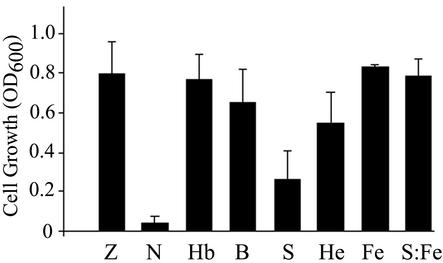
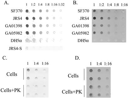
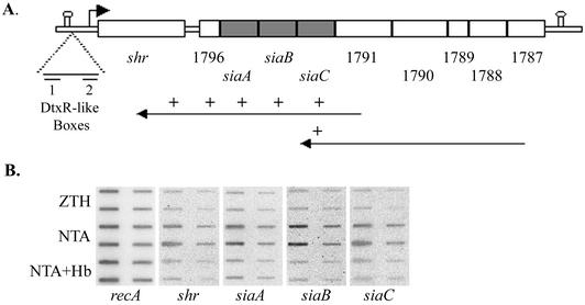
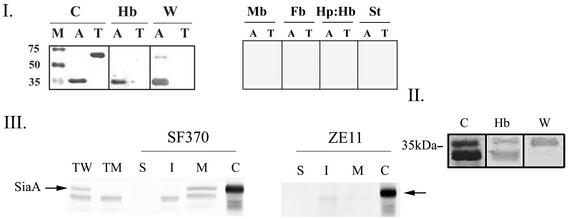
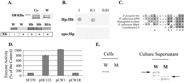
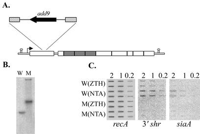
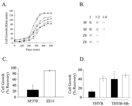
The hemolytic Streptococcus pyogenes can use a variety of heme compounds as an iron source. In this study, we investigate hemoprotein utilization by S. pyogenes. We demonstrate that surface proteins contribute to the binding of hemoproteins to S. pyogenes. We identify an ABC transporter from the iron complex family named sia for streptococcal iron acquisition, which consists of a lipoprotein (siaA), membrane permease (siaB), and ATPase (siaC). The sia transporter is part of a highly conserved, iron regulated, 10-gene operon. SiaA, which was localized to the cell membrane, could specifically bind hemoglobin. The operon's first gene encodes a novel bacterial protein that bound hemoglobin, myoglobin, heme-albumin, and hemoglobin-haptoglobin (but not apo-haptoglobin) and therefore was named Shr, for streptococcal hemoprotein receptor. PhoZ fusion and Western blot analysis showed that Shr has a leader peptide and is found in both membrane-bound and soluble forms. An M1 SF370 strain with a polar mutation in shr was more resistant to streptonigrin and hydrogen peroxide, suggesting decreased iron uptake. The addition of hemoglobin to the culture medium increased cell resistance to hydrogen peroxide in SF370 but not in the mutant, implying the sia operon may be involved in hemoglobin-dependent resistance to oxidative stress. The shr mutant demonstrated reduced hemoglobin binding, though cell growth in iron-depleted medium supplemented with hemoglobin, whole blood, or ferric citrate was not affected, suggesting additional systems are involved in hemoglobin utilization. SiaA and Shr are the first hemoprotein receptors identified in S. pyogenes; their possible role in iron capture is discussed.


Figures

FIG. 1.
FIG. 1.
Use of host iron sources by S. pyogenes strain SF370. Cells were used to inoculate (1:1,000 dilution from glycerol stocks) fresh ZTH medium (Z), ZTH medium containing 10 mM NTA (N), ZTH-NTA medium supplemented with 22 μM hemoglobin (Hb), 0.125% whole sheep blood (B), 0.125% horse serum (S), 6 μM heme-BSA (He), 10 mM ferric citrate (Fe), or 0.125% serum and 10 mM ferric citrate (S:Fe). Cell growth is expressed as the culture OD600 after overnight incubation (17 h). The results are from at least four separate experiments; the error bars indicate the standard deviation.
FIG. 2.
FIG. 2.
Hemoglobin and heme binding to S. pyogenes surface. (A) Binding to human hemoglobin by S. pyogenes isolates. Cell suspensions at an OD600 of 1 were immobilized onto a nitrocellulose membrane from a serial dilution. Binding to biotinylated hemoglobin was detected with streptavidin-conjugated alkaline phosphatase. Binding to S. pyogenes strains SF370, JRS4, GA01398, and GA05982 and to E. coli DH5α is shown. JRS4-S represents JRS4 cells treated only with streptavidin. (B) Heme binding to S. pyogenes surface. The results here are the same as in panel A except that the membranes were stained with the chromogenic substrate DMB. (C) Surface proteins mediate hemoglobin binding to S. pyogenes. SF370 cells were incubated for an hour in buffer (cells) or were treated with proteinase K (cells+PK) prior to membrane immobilization in duplicates. Hemoglobin binding was assayed as in panel A. (D) Nonprotein constituent(s) mediates most of the heme binding to S. pyogenes. The results here are the same as in panel C except that the membrane was stained with DMB.
FIG. 3.
FIG. 3.
S. pyogenes iron acquisition (sia) locus. (A) The sia operon consists of 10 genes flanked by predicted transcription terminators (represented by stem-loops); the numbers below indicate the SF370 gene number, and the small arrow shows the location of the putative shr promoter. The long arrows represent the cDNAs produced for the RT-PCR analysis and the “+” symbols represent sia sequences found on the cDNAs. (B) RNA slot blots with RNA extracted from mid-log-phase SF370 grown in ZTH, ZTH and NTA (NTA), or ZTH with NTA and hemoglobin (NTA+Hb). We applied 2 and 0.4 μg of RNA from each sample to the membrane in duplicates.
FIG. 4.
FIG. 4.
(I) Purified recombinant SiaA (rSiaA) binds hemoglobin in vitro. Purified rSiaA (His-Xpress-SiaA, A) and human transferrin (T) were fractionated by SDS-PAGE under seminative conditions. A Coomassie blue-stained gel (C), Western blot analysis with anti-express antibodies (W, 1:5,000 dilution), and binding blots to biotinylated hemoglobin (Hb), myoglobin (Mb), fibrinogen (Fb), haptoglobin-hemoglobin (Hp:Hb), or streptavidin (St)-conjugated alkaline phosphatase are presented. M, molecular weight marker. (II) rSiaA binding to hemoglobin is independent of the His-Xpress epitope. The results here are the same as in panel A, except the rSiaA was digested with enterokinase prior to analysis. Both the digested and the undigested rSiaA can be seen in the Coomassie blue stain and the hemoglobin blot. (III) SiaA is associated with the S. pyogenes envelope. Total protein extracts prepared from the wild-type SF370 (TW) and the mutant ZE11 (TM) were analyzed by Western blot analysis with anti-rSiaA serum (1:5,000 dilution). Proteins from various cell fractions of SF370 and ZE11 strains are included: S, surface proteins; I, intracellular proteins; and M, membrane proteins. Total protein extracts from whole S. pyogenes SF370/pCPSiaA cells expressing the native SiaA from P23 promoter is included as a positive control (C). The arrows indicate SiaA.
FIG. 5.
FIG. 5.
(A) Purified rShr (His-Xpress-Shr) was fractionated by SDS-PAGE under seminative conditions. Coomassie blue-stained gel (Co), Western blot analysis with anti-Xpress antibodies (W, 1:5,000 dilution) and binding blots to biotinylated hemoglobin (Hb), myoglobin (Mb), and heme-BSA (BSA) are shown. The “+” sign indicates digestion of rShr with enterokinase prior to analysis. (B) rShr was spotted onto a membrane (1, 0.1, and 0.01 μg/spot) and reacted with biotinylated haptoglobin-hemoglobin (Hp:Hb) or apo-haptoglobin (apo:Hp). (C) Shr heme-binding motif. (D) PhoZ enzyme activity in cell-free culture supernatant of S. pyogenes harboring Shr-PhoZ fusion. Enzyme activity is expressed as a percentage of the background activity measured in SF370. Activity in cultures of cells harboring PhoZ expressed with its native leader peptide (pDC123, positive control), Shr-PhoZ fusion (pCW1), and Shr-PhoZ fusion in a reverse orientation (pCW1R, negative control) are shown. The mean and standard deviation of at least two independent experiments, each conducted in duplicates, are shown. (E) Western blot analysis of proteins prepared from SF370 (W) and ZE11 (M) strains with anti-rShr serum (1:5,000 dilution). Proteins prepared from cells and cell-free culture supernatant are included. The arrows point to the full-length Shr in wild-type and mutant strains.
FIG. 6.
FIG. 6.
Construction and analysis of an insertion duplication mutant in the sia operon. (A) Schematic presentation of the shr::pCB2 mutation. The insertion site and the structure of plasmid pCB2 are indicated. The dashed boxes indicate the internal shr fragment cloned into pCB2. (B) Southern blot analysis. Chromosomal DNA of the parent strain SF370 (W) and the mutant strain ZE11 (M) was digested with BglII and hybridized with a [α-32P]dATP-labeled probe specific to the shr gene. (C) RNA slot blot analysis of sia genes in both the wild-type (W) and the mutant (M) strains. The same as in Fig. 3C, RNA was extracted from mid-log-phase SF370 grown in complete medium (ZTH) or iron-restricted medium (NTA).
FIG. 7.
FIG. 7.
Analysis of strain ZE11. (A) Growth of the wild-type strain SF370 (solid shapes) and the mutant strain ZE11 (open shapes). Cells were used to inoculate (1:100 dilution) fresh ZTH medium (diamonds), NTA containing medium (squares), and NTA-medium supplemented with 8 μM hemoglobin (circles). (B) Binding of biotinylated hemoglobin to cells of SF370 (SF) and ZE11 (ZE). The hemoglobin-binding assay was done as described in Fig. 2A. (C) Sensitivity to streptonigrin of the wild-type and the mutant strains. Cells were used to inoculate (1:1,000 dilution) fresh ZTH medium and medium containing 1.6 μM streptonigrin. The culture optical density (OD600) after overnight incubation (17 h) was determined. The data are presented as the percent growth of each strain in ZTH. The results are from seven independent experiments; the error bars indicate the standard deviation (P < 0.01, t test). (D) Sensitivity of SF370 and ZE11 to hydrogen peroxide. Overnight cultures were used to inoculate THYB or THYB containing 15 μM hemoglobin. Cells in mid-log phase were challenged with 5 mM hydrogen peroxide for 30 min. Viable counts were determined by plating samples taken before and after the challenge, and cell recovery was expressed as the percentage. The data represent results obtained from at least three experiments and plating was done in triplicate. Error bars represent the standard error of the mean (P < 0.01 for SF370 and ZE11 in THYB and P < 0.02 for SF370 in THYB and SF370 in THYB+Hb according to the Student t test).

References

    1. Al-Tawfiq, J. A., K. R. Fortney, B. P. Katz, A. F. Hood, C. Elkins, and S. M. Spinola. 2000. An isogenic hemoglobin receptor-deficient mutant of Haemophilus ducreyi is attenuated in the human model of experimental infection. J. Infect. Dis. 181:1049-1054. - PubMed
    1. Altman, P. L., and D. D. Katz. 1961. Blood and other body fluids. Federation of American Societies for Experimental Biology, Washington, D.C.
    1. Bracken, C. S., M. T. Baer, A. Abdur-Rashid, W. Helms, and I. Stojiljkovic. 1999. Use of heme-protein complexes by the Yersinia enterocolitica HemR receptor: histidine residues are essential for receptor function. J. Bacteriol. 181:6063-6072. - PMC - PubMed
    1. Brown, J. S., S. M. Gilliland, and D. W. Holden. 2001. A Streptococcus pneumoniae pathogenicity island encoding an ABC transporter involved in iron uptake and virulence. Mol. Microbiol. 40:572-585. - PubMed
    1. Brown, J. S., A. D. Ogunniyi, M. C. Woodrow, D. W. Holden, and J. C. Paton. 2001. Immunization with components of two iron uptake ABC transporters protects mice against systemic Streptococcus pneumoniae infection. Infect. Immun. 69:6702-6706. - PMC - PubMed

Publication types