

Structural characterization of the N terminus of IpaC from Shigella flexneri
- PMID: 12595440
- PMCID: PMC148864
- DOI: 10.1128/IAI.71.3.1255-1264.2003
Structural characterization of the N terminus of IpaC from Shigella flexneri
Abstract
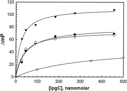
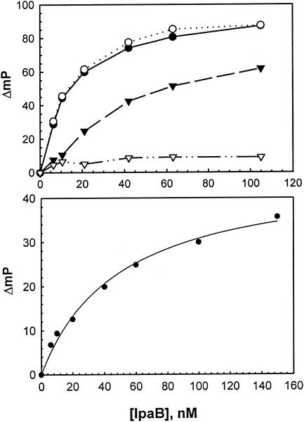
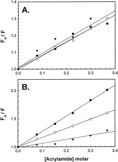
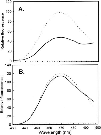
The primary effector for Shigella invasion of epithelial cells is IpaC, which is secreted via a type III secretion system. We recently reported that the IpaC N terminus is required for type III secretion and possibly other functions. In this study, mutagenesis was used to identify an N-terminal secretion signal and to determine the functional importance of the rest of the IpaC N terminus. The 15 N-terminal amino acids target IpaC for secretion by Shigella flexneri, and placing additional amino acids at the N terminus does not interfere with IpaC secretion. Furthermore, amino acid sequences with no relationship to the native IpaC secretion signal can also direct its secretion. Deletions introduced beyond amino acid 20 have no effect on secretion and do not adversely affect IpaC function in vivo until they extend beyond residue 50, at which point invasion function is completely eliminated. Deletions introduced at amino acid 100 and extending toward the N terminus reduce IpaC's invasion function but do not eliminate it until they extend to the N-terminal side of residue 80, indicating that a region from amino acid 50 to 80 is critical for IpaC invasion function. To explore this further, the ability of an IpaC N-terminal peptide to associate in vitro with its translocon partner IpaB and its chaperone IpgC was studied. The N-terminal peptide binds tightly to IpaB, but the IpaC central hydrophobic region also appears to participate in this binding. The N-terminal peptide also associates with the chaperone IpgC and IpaB is competitive for this interaction. Based on additional biophysical data, we propose that a region between amino acids 50 and 80 is required for chaperone binding, and that the IpaB binding domain is located downstream from, and possibly overlapping, this region. From these data, we propose that the secretion signal, chaperone binding region, and IpaB binding domain are located at the IpaC N terminus and are essential for presentation of IpaC to host cells during bacterial entry; however, IpaC effector activity may be located elsewhere.
Figures
Similar articles
-
The C-terminus of IpaC is required for effector activities related to Shigella invasion of host cells.Microb Pathog. 2008 Oct;45(4):282-9. doi: 10.1016/j.micpath.2008.06.003. Epub 2008 Jul 4. Microb Pathog. 2008. PMID: 18656530 Free PMC article.
-
Preparation and characterization of translocator/chaperone complexes and their component proteins from Shigella flexneri.Biochemistry. 2007 Jul 10;46(27):8128-37. doi: 10.1021/bi700099c. Epub 2007 Jun 16. Biochemistry. 2007. PMID: 17571858
-
IpaD of Shigella flexneri is independently required for regulation of Ipa protein secretion and efficient insertion of IpaB and IpaC into host membranes.Infect Immun. 2005 Mar;73(3):1432-40. doi: 10.1128/IAI.73.3.1432-1440.2005. Infect Immun. 2005. PMID: 15731041 Free PMC article.
-
Molecular and Cellular Mechanisms of Shigella flexneri Dissemination.Front Cell Infect Microbiol. 2016 Mar 11;6:29. doi: 10.3389/fcimb.2016.00029. eCollection 2016. Front Cell Infect Microbiol. 2016. PMID: 27014639 Free PMC article. Review.
-
Bacterial signals and cell responses during Shigella entry into epithelial cells.Cell Microbiol. 2000 Jun;2(3):187-93. doi: 10.1046/j.1462-5822.2000.00046.x. Cell Microbiol. 2000. PMID: 11207575 Review.
Cited by
-
N-terminus of IpaB provides a potential anchor to the Shigella type III secretion system tip complex protein IpaD.Biochemistry. 2013 Dec 10;52(49):8790-9. doi: 10.1021/bi400755f. Epub 2013 Nov 20. Biochemistry. 2013. PMID: 24236510 Free PMC article.
-
Liposomes recruit IpaC to the Shigella flexneri type III secretion apparatus needle as a final step in secretion induction.Infect Immun. 2009 Jul;77(7):2754-61. doi: 10.1128/IAI.00190-09. Epub 2009 May 11. Infect Immun. 2009. PMID: 19433542 Free PMC article.
-
Protein export according to schedule: architecture, assembly, and regulation of type III secretion systems from plant- and animal-pathogenic bacteria.Microbiol Mol Biol Rev. 2012 Jun;76(2):262-310. doi: 10.1128/MMBR.05017-11. Microbiol Mol Biol Rev. 2012. PMID: 22688814 Free PMC article. Review.
-
Conformational stability and differential structural analysis of LcrV, PcrV, BipD, and SipD from type III secretion systems.Protein Sci. 2007 Apr;16(4):704-14. doi: 10.1110/ps.062645007. Epub 2007 Feb 27. Protein Sci. 2007. PMID: 17327391 Free PMC article.
-
Combination of two separate binding domains defines stoichiometry between type III secretion system chaperone IpgC and translocator protein IpaB.J Biol Chem. 2010 Dec 17;285(51):39965-75. doi: 10.1074/jbc.M110.135616. Epub 2010 Oct 11. J Biol Chem. 2010. PMID: 20937829 Free PMC article.
References
-
- Barbieri, J. T., M. J. Riese, and K. Aktories. 2002. Bacterial toxins that modify the actin cytoskeleton. Annu. Rev. Cell Dev. Biol. 18:315-344. - PubMed
-
- Bennett, J. C., and C. Hughes. 2000. From flagellum assembly to virulence: the extended family of type III export chaperones. Trends Microbiol. 8:202-204. - PubMed
-
- Birtalan, S. C., R. M. Phillips, and P. Ghosh. 2002. Three-dimensional secretion signals in chaperone-effector complexes of bacterial pathogens. Mol. Cell 9:971-980. - PubMed
Publication types
MeSH terms
Substances
Grants and funding
LinkOut - more resources
Full Text Sources
Other Literature Sources
