. 2003 Mar;71(3):1370-8.
doi: 10.1128/IAI.71.3.1370-1378.2003.

Primed peritoneal B lymphocytes are sufficient to transfer protection against Brugia pahangi infection in mice

Affiliations

Primed peritoneal B lymphocytes are sufficient to transfer protection against Brugia pahangi infection in mice

Natalia Paciorkowski et al. Infect Immun. 2003 Mar.

Abstract

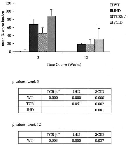
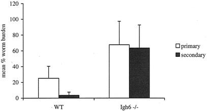
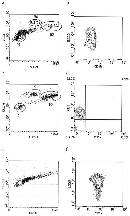
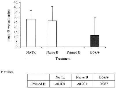
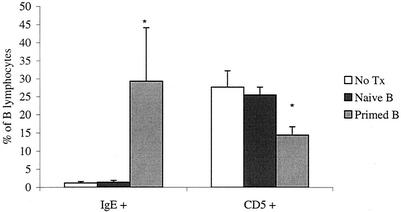
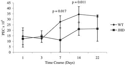
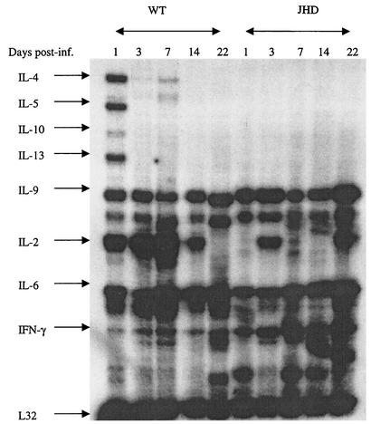
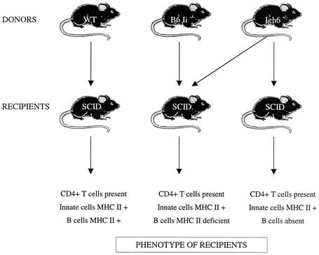
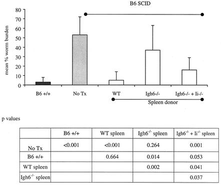
Lymphatic filariasis is a tropical disease caused by the nematode parasites Wuchereria bancrofti and Brugia malayi. Whereas the protective potential of T lymphocytes in filarial infection is well documented, investigation of the role of B lymphocytes in antifilarial immunity has been neglected. In this communication, we examine the role of B lymphocytes in antifilarial immunity, using Brugia pahangi infections in the murine peritoneal cavity as a model. We find that B lymphocytes are required for clearance of primary and challenge infections with B. pahangi third-stage larvae (L3). We assessed the protective potential of peritoneal B lymphocytes by adoptive transfer experiments. Primed but not naïve peritoneal B cells from wild-type mice that had been immunized with B. pahangi L3 protected athymic recipients from challenge infection. We evaluated possible mechanisms by which B cells mediate protection. Comparisons of cytokine mRNA expression between B-lymphocyte-deficient and immunocompetent mice following B. pahangi infection suggest that B cells are required for the early production of Th2-type cytokines by peritoneal cells. In addition, B-cell-deficient mice demonstrate a defect in inflammatory cell recruitment to the peritoneal cavity following B. pahangi infection. The data demonstrate a critical role of B lymphocytes in antifilarial immunity in naïve mice and in the memory response in primed mice.


Figures

FIG. 1.
FIG. 1.
B. pahangi recoveries from BALB/c+/+, BALB/c TCRβ−/−, BALB/c JHD, and BALB/c SCID mice. BALB/c+/+, BALB/c TCRβ−/−, BALB/c JHD, and BALB/c SCID mice were infected with 50 B. pahangi L3. Worm recoveries were quantitated as described in the text. Each bar represents the average from five mice. Microfilariae were identified by microscopic analysis of the peritoneal lavage.
FIG. 2.
FIG. 2.
Comparison between B. pahangi recoveries following primary or challenge infection in B6+/+ (WT) and B6 Igh6−/− mice. B6+/+ and B6 Igh6−/− mice were immunized with 50 B. pahangi L3 i.p. Two months following immunization, these groups and nonmanipulated control groups received a challenge infection with 50 B. pahangi L3. Worm recoveries were quantitated at 2 weeks postinfection. The bars represent the average from eight mice for all the groups except the Igh6−/− group that received only challenge infection, which had seven mice. P is <0.001 for wild-type mice.
FIG. 3.
FIG. 3.
Donor PECs used for reconstitution of B6 NUDE mice. (a to d) Twenty B6+/+ mice were immunized with 25 B. pahangi L3. PECs were collected at 3 weeks following immunization and B cells were purified. Panels a and b show the positively selected fraction. Gates R1, R3, and R4 in panel a demarcate small lymphocytes, macrophages, and eosinophils, respectively. Panels c and d show the B-cell-depleted flowthrough fraction. Panel d is gated on small lymphocytes (R1). Cells were stained with anti-CD19-FITC and B220-PE, and anti-CD3-PE. (e and f) PECs were collected from 35 naïve B6+/+ mice. B lymphocytes were purified using the same protocol and reagents as described for primed donor cells. Both panels show positively selected CD19+ peritoneal cells. Cells were stained with anti-CD19-FITC and B220-PE.
FIG. 4.
FIG. 4.
B. pahangi recoveries from NUDE mice reconstituted with purified wild-type peritoneal B lymphocytes. B6 NUDE mice were reconstituted with 5 × 10⁶ naïve or primed peritoneal B lymphocytes via i.p. injection. One week later, both reconstituted cohorts, a group of nonmanipulated B6 NUDE mice (No Tx), and a group of B6+/+ mice were infected with 50 B. pahangi L3 intraperitoneally. All mice were necropsied at 2 weeks postinfection and worm burdens were quantitated. n = 10 for the No Tx and primed B groups; n = 9 for the naïve B group; n = 5 for the B6+/+ group.
FIG. 5.
FIG. 5.
Proportions of IgE+ and CD5+ B lymphocytes in the peritoneal cavity of nude mice reconstituted with naïve or primed peritoneal B cells. PECs were gated on lymphocytes and analyzed using anti-IgE-FITC and anti-CD19-biotin or anti-CD19-FITC and anti-CD5-PE. Each bar represents the average from 10 (primed B and no treatment [No Tx] groups) or 9 (naïve B group) mice. The values from the primed B group for both cell types were significantly different from the other groups (P < 0.000).
FIG. 6.
FIG. 6.
Comparisons of PEC accumulation following B. pahangi infection between B6+/+ (WT) and B6 JHD mice. Groups of 25 B6+/+ and 25 B6 JHD mice were infected with 50 B. pahangi L3. Cohorts of five mice from each group were necropsied at different time points postinfection. PECs were recovered and counted by using a hemocytometer.
FIG. 7.
FIG. 7.
Cytokine expression by PECs from B6+/+ (WT) and B6 JHD mice infected with B. pahangi. Groups of 25 B6+/+ mice and 25 B6 JHD mice were infected with 50 B. pahangi L3. Five mice from each cohort were sacrificed on days 1, 3, 7, 14, and 22 postinfection. PECs were collected from each group, pooled, and used for RNA preparation. Forty micrograms of RNA from each group was used for an RNase protection assay using the mCK-1 probe. Each lane shows cytokine expression from five mice.
FIG. 8.
FIG. 8.
Experimental design for the investigation of the requirement for the antigen-presenting function of B lymphocytes.
FIG. 9.
FIG. 9.
B. pahangi recoveries from B6 SCID mice reconstituted with different antigen-presenting cell types. Ten B6 SCID mice were reconstituted with 2 × 10⁷ spleen cells from B6+/+ mice (WT), 10 B6 SCID mice received 107 spleen cells from B6 Igh6−/− mice, and 10 B6 SCID mice received an equal mixture of spleen cells from B6 Igh6−/− mice and Ii−/− mice (107 cells from each donor per recipient). All cells were injected i.p. One day later the reconstituted mice, B6+/+ mice (n = 5), and nonmanipulated B6 SCID mice (No Tx; n = 5) were infected with 50 B. pahangi L3. Mice were necropsied at 2 weeks postinfection and worm burdens were quantitated as described in the text.
FIG. 10.
FIG. 10.
Transfer of primed serum to CBA/N mice. Thirty-five CBA/Ca mice were immunized with 40 B. pahangi L3. Mice were bled by cardiac puncture at 3 weeks postinfection; serum was collected by centrifugation and pooled. Five CBA/Ca mice were infected with 45 B. pahangi L3 and injected i.p. with 240 μl (the volume-equivalent of serum that we usually recover from a single mouse) of pooled serum on days 0, 2, 4, 6, 8, 10, and 12 postinfection. The control groups of CBA/N mice received no treatment (No Tx) or were injected with 240 μl of pooled serum from B. pahangi-infected B6 Igh6−/− mice (n = 5 for each group). All mice were necropsied on day 14 postinfection and worm burdens were quantitated.

References

    1. Al-Qaoud, K. M., E. Pearlman, T. Hartung, J. Klukowski, B. Fleischer, and A. Hoerauf. 2000. A new mechanism for IL-5-dependent helminth control: neutrophil accumulation and neutrophil-mediated worm encapsulation in murine filariasis are abolished in the absence of IL-5. Int. Immunol. 12:899-908. - PubMed
    1. Anonymous. 2001. Lymphatic filariasis. Wkly. Epidemiol. Rec. 76:149-154. - PubMed
    1. Arulanandam, B. P., J. M. Lynch, D. E. Briles, S. Hollingshead, and D. W. Metzger. 2001. Intranasal vaccination with pneumococcal surface protein A and interleukin-12 augments antibody-mediated opsonization and protective immunity against Streptococcus pneumoniae infection. Infect. Immun. 69:6718-6724. - PMC - PubMed
    1. Bancroft, A. J., R. K. Grencis, K. J. Else, and E. Devaney. 1994. The role of CD4 cells in protective immunity to Brugia pahangi. Parasite Immunol. 16:385-387. - PubMed
    1. Bikah, G., J. Carey, J. R. Ciallella, A. Tarakhovsky, and S. Bondada. 1996. CD5-mediated negative regulation of antigen receptor-induced growth signals in B-1 B cells. Science 274:1906-1909. - PubMed

LinkOut - more resources