

Intravenous mouse infection model for studying the pathology of Enterococcus faecalis infections
- PMID: 12595461
- PMCID: PMC148842
- DOI: 10.1128/IAI.71.3.1434-1441.2003
Intravenous mouse infection model for studying the pathology of Enterococcus faecalis infections
Abstract
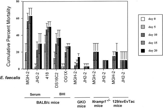
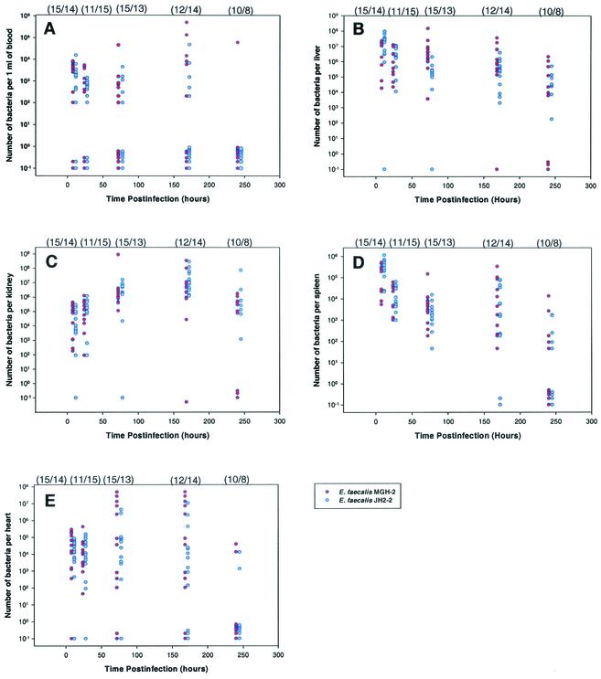
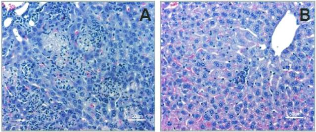
An intravenous mouse infection model was used to compare the virulence of Enterococcus faecalis strains, to study bacterial localization and organ histopathology, and to examine the effects of Nramp1 and gamma interferon (IFN-gamma) on the course of infection. Infection of BALB/c mice with 5 x 10(8) CFU of E. faecalis JH2-2, MGH-2, 418, DS16C2, or OG1X revealed the following virulence ranking (from highest to lowest): MGH-2, 418, DS16C2, JH2-2, and OG1X. Discernible differences in the number of MGH-2 and JH2-2 bacteria were observed at 7 days (168 h) in the blood (P = 0.037), at 72 h in the liver (P = 0.002), and at 8 h in the spleen (P = 0.036). At these time points, the number of MGH-2 bacteria was higher in the blood and liver while the number of JH2-2 bacteria was higher in the spleen. At 72 h, livers from MGH-2-infected mice had higher numbers of coalescing aggregates of leukocytes and a greater degree of caseous necrosis than those from JH2-2-infected mice. These results indicate a correlation between the virulence of the E. faecalis strain, the number of bacteria in the liver, and the degree of histopathology of the liver at 72 h postinfection. IFN-gamma was important in E. faecalis infection, since IFN-gamma gene knockout mice had reduced mortality and massive coagulative necrosis was observed in wild-type mice. The contribution of Nramp1 was unclear, since Nramp1(-/-) mice and the respective control mice were innately resistant to E. faecalis. The mortality of mice in this model is probably due to induction of cytokine release and massive coagulative necrosis.
Figures
Similar articles
-
In vivo assessment of growth and virulence gene expression during commensal and pathogenic lifestyles of luxABCDE-tagged Enterococcus faecalis strains in murine gastrointestinal and intravenous infection models.Appl Environ Microbiol. 2013 Jul;79(13):3986-97. doi: 10.1128/AEM.00831-13. Epub 2013 Apr 19. Appl Environ Microbiol. 2013. PMID: 23603680 Free PMC article.
-
Survival of Enterococcus faecalis in mouse peritoneal macrophages.Infect Immun. 1999 May;67(5):2160-5. doi: 10.1128/IAI.67.5.2160-2165.1999. Infect Immun. 1999. PMID: 10225869 Free PMC article.
-
Contribution of secreted proteases to the pathogenesis of postoperative Enterococcus faecalis endophthalmitis.J Cataract Refract Surg. 2008 Oct;34(10):1776-84. doi: 10.1016/j.jcrs.2008.06.033. J Cataract Refract Surg. 2008. PMID: 18812133
-
A genomic virulence reference map of Enterococcus faecalis reveals an important contribution of phage03-like elements in nosocomial genetic lineages to pathogenicity in a Caenorhabditis elegans infection model.Infect Immun. 2015 May;83(5):2156-67. doi: 10.1128/IAI.02801-14. Epub 2015 Mar 16. Infect Immun. 2015. PMID: 25776747 Free PMC article.
-
Molecular Mechanism of Quorum-Sensing in Enterococcus faecalis: Its Role in Virulence and Therapeutic Approaches.Int J Mol Sci. 2017 May 3;18(5):960. doi: 10.3390/ijms18050960. Int J Mol Sci. 2017. PMID: 28467378 Free PMC article. Review.
Cited by
-
Redox balance via lactate dehydrogenase is important for multiple stress resistance and virulence in Enterococcus faecalis.Infect Immun. 2013 Aug;81(8):2662-8. doi: 10.1128/IAI.01299-12. Epub 2013 May 6. Infect Immun. 2013. PMID: 23649090 Free PMC article.
-
Bioactivity Studies of β-Lactam Derived Polycyclic Fused Pyrroli-Dine/Pyrrolizidine Derivatives in Dentistry: In Vitro, In Vivo and In Silico Studies.PLoS One. 2015 Jul 17;10(7):e0131433. doi: 10.1371/journal.pone.0131433. eCollection 2015. PLoS One. 2015. PMID: 26185985 Free PMC article.
-
The transcriptome of the nosocomial pathogen Enterococcus faecalis V583 reveals adaptive responses to growth in blood.PLoS One. 2009 Nov 4;4(11):e7660. doi: 10.1371/journal.pone.0007660. PLoS One. 2009. PMID: 19888459 Free PMC article.
-
Histopathological changes induced in an animal model by potentially pathogenic Enterococcus faecalis strains recovered from ready-to-eat food outlets in Osun State, Nigeria.Infect Drug Resist. 2015 Jun 26;8:181-7. doi: 10.2147/IDR.S61381. eCollection 2015. Infect Drug Resist. 2015. PMID: 26170700 Free PMC article.
-
In vivo assessment of growth and virulence gene expression during commensal and pathogenic lifestyles of luxABCDE-tagged Enterococcus faecalis strains in murine gastrointestinal and intravenous infection models.Appl Environ Microbiol. 2013 Jul;79(13):3986-97. doi: 10.1128/AEM.00831-13. Epub 2013 Apr 19. Appl Environ Microbiol. 2013. PMID: 23603680 Free PMC article.
References
-
- Antalek, M. D., J. M. Mylotte, A. J. Lesse, and J. A. Sellick, Jr. 1995. Clinical and molecular epidemiology of Enterococcus faecalis bacteremia, with special reference to strains with high-level resistance to gentamicin. Clin. Infect. Dis. 20:103-109. - PubMed
-
- Caballero-Granado, F. J., B. Becerril, J. M. Cisneros, L. Cuberos, I. Moreno, and J. Pachon. 2001. Case-control study of risk factors for the development of enterococcal bacteremia. Eur. J. Clin. Microbiol. Infect. Dis. 20:83-90. - PubMed
-
- Caballero-Granado, F. J., B. Becerril, L. Cuberos, M. Bernabeu, J. M. Cisneros, and J. Pachon. 2001. Attributable mortality rate and duration of hospital stay associated with enterococcal bacteremia. Clin. Infect. Dis. 32:587-594. - PubMed
-
- Caballero-Granado, F. J., J. M. Cisneros, R. Luque, M. Torres-Tortosa, F. Gamboa, F. Diez, J. L. Villanueva, R. Perez-Cano, J. Pasquau, D. Merino, A. Menchero, D. Mora, M. A. Lopez-Ruz, A. Vergara, et al. 1998. Comparative study of bacteremias caused by Enterococcus spp. with and without high-level resistance to gentamicin. J. Clin. Microbiol. 36:520-525. - PMC - PubMed
Publication types
MeSH terms
Substances
Grants and funding
LinkOut - more resources
Full Text Sources
Other Literature Sources
