. 2003 Mar;71(3):1442-52.
doi: 10.1128/IAI.71.3.1442-1452.2003.

Role of the NF-kappaB signaling pathway and kappaB cis-regulatory elements on the IRF-1 and iNOS promoter regions in mycobacterial lipoarabinomannan induction of nitric oxide

Affiliations

Role of the NF-kappaB signaling pathway and kappaB cis-regulatory elements on the IRF-1 and iNOS promoter regions in mycobacterial lipoarabinomannan induction of nitric oxide

Kristin R Morris et al. Infect Immun. 2003 Mar.

Abstract

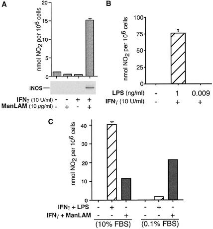
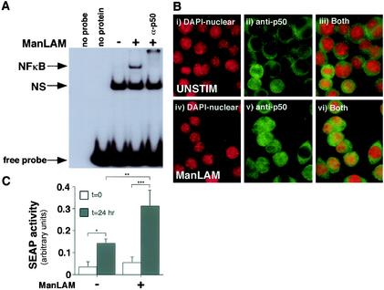
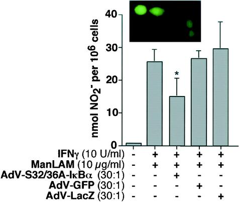
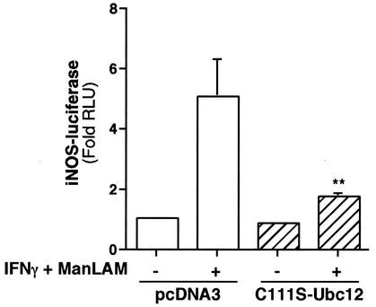
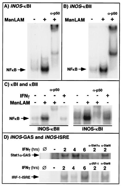
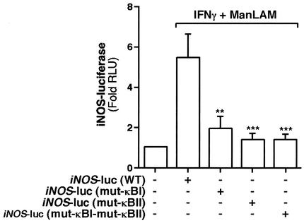
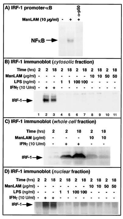
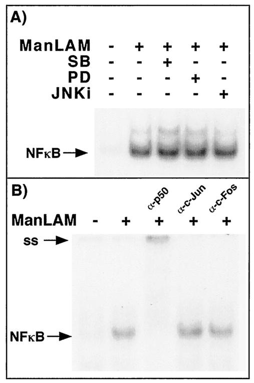
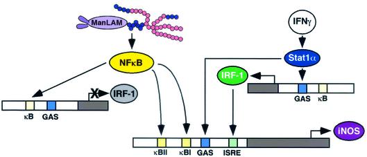
Nitric oxide (NO(.)) produced by inducible nitric oxide synthase (iNOS) is an important host defense molecule against Mycobacterium tuberculosis in mononuclear phagocytes. The objective of this study was to determine the role of the IkappaBalpha kinase-nuclear factor kappaB (IKK-NF-kappaB) signaling pathway in the induction of iNOS and NO(.) by a mycobacterial cell wall lipoglycan known as mannose-capped lipoarabinomannan (ManLAM) in mouse macrophages costimulated with gamma interferon (IFN-gamma). NF-kappaB was activated by ManLAM as shown by electrophoretic mobility shift assay, by immunofluorescence of translocated NF-kappaB in intact cells, and by a reporter gene driven by four NF-kappaB-binding elements. Transduction of an IkappaBalpha mutant (Ser32/36Ala) significantly inhibited NO(.) expression induced by IFN-gamma plus ManLAM. An activated SCF complex, a heterotetramer (Skp1, Cul-1, beta-TrCP [F-box protein], and ROC1) involved with ubiquitination, is also required for iNOS-NO(.) induction. Two NF-kappaB-binding sites (kappaBI and kappaBII) present on the 5'-flanking region of the iNOS promoter bound ManLAM-induced NF-kappaB similarly. By use of reporter constructs in which one or both sites are mutated, both NF-kappaB-binding positions were essential in iNOS induction by IFN-gamma plus ManLAM. IFN-gamma-induced activation of the IRF-1 transcriptional complex is a necessary component in host defense against tuberculosis. Although the 5'-flanking region of the IRF-1 promoter contains an NF-kappaB-binding site and ManLAM-induced NF-kappaB also binds to this site, ManLAM was unable to induce IRF-1 expression. The influence of mitogen-activated protein kinases on IFN-gamma plus ManLAM induction of iNOS-NO(.) is not due to any effects on ManLAM induction of NF-kappaB.


Figures

FIG. 1.
FIG. 1.
Effects of IFN-γ plus ManLAM induction of NO· are not due to LPS contamination. (A) (Top) RAW264.7γNO(−) cells were either left unstimulated or stimulated with either 10 U of IFN-γ/ml, 10 μg of ManLAM/ml, or both for 18 h, followed by an assay for NO2 in the supernatant. (Bottom) Corresponding immunoblot of the whole-cell lysate for iNOS protein. (B) RAW264.7γNO(−) cells were stimulated with 10 U of IFN-γ/ml plus 1 ng of LPS/ml or 10 U of IFN-γ/ml plus 0.009 ng of LPS/ml (the latter being the concentration of LPS contained in 10 μg of ManLAM/ml), followed by an NO2 assay. (C) RAW 264.7γNO(−) cells were stimulated with either IFN-γ (10 U/ml) plus LPS (1 ng/ml) or IFN-γ (10 U/ml) plus ManLAM (10 μg/ml) in the presence of either 10% FBS or 0.1% FBS in the culture medium. Data shown are means ± SD from three independent experiments.
FIG. 2.
FIG. 2.
ManLAM activates functional NF-κB. (A) RAW 264.7γNO(−) cells were stimulated with ManLAM (10 μg/ml) for 30 min, and nuclear proteins were isolated, followed by an EMSA with a 32P-labeled NF-κB consensus oligonucleotide. Compared to unstimulated cells, ManLAM induced NF-κB activation. The specificity of the NF-κB complex was verified by coincubation of the nuclear extract-oligonucleotide mixture with an anti-p50 antibody (α-p50). The nonspecific (NS) band was present in both unstimulated and ManLAM-stimulated cells and did not supershift with the anti-p50 antibody. (B) Unstimulated and ManLAM-stimulated RAW 264.7γNO(−) cells were stained with DAPI for nuclear detection and immunostained with an anti-p50-NF-κB antibody. The cells were then viewed under a fluorescent microscope at selective wavelengths to detect nuclear staining (i and iv), p50-NF-κB staining (ii and v), or both (iii and vi). After counting of 200 cells in random fields, the percentage of cells with positive nuclear staining for p50-NF-κB was 12.5% for unstimulated cells and 46.3% for ManLAM-stimulated cells. (C) RAW 264.7γNO(−) cells were transfected with 2 μg of the κB-SEAP plasmid. At 0 and 24 h after stimulation with) ManLAM (10 μg/ml), the supernatant was assayed for alkaline phosphatase activity (*, P < 0.05; **, P < 0.01; ***, P < 0.001). Data shown in panels A and B are representative of three independent experiments. Data shown in panel C are means ± SD from three independent experiments.
FIG. 3.
FIG. 3.
Mutant IκBα inhibits NO· expression. RAW 264.7γNO(−) were transduced with AdV-S32/36A-IκBα, AdV-GFP, or AdV-LacZ at an MOI ratio of 30:1. After 6 h of incubation, the cells were stimulated with IFN-γ (10 U/ml) plus ManLAM (10 μg/ml) for 18 h, followed by an assay of the cell supernatant for NO2 (*, P < 0.05). Data are means ± SD from three independent experiments. (Inset) RAW 264.7γNO(−) cells transduced with AdV-GFP and viewed under a fluorescent microscope. Data are means ± SD from three independent experiments.
FIG. 4.
FIG. 4.
NEDD8 conjugation to Cul-1 is required for IFN-γ plus ManLAM induction of iNOS promoter activity. RAW 264.7γNO(−) cells were cotransfected with 0.3 μg of the iNOS-luc plasmid and 2 μg of the mutant Ubc12 (C111S-Ubc12) plasmid or the empty vector pcDNA3, followed by stimulation with IFN-γ (10 U/ml) plus ManLAM (10 μg/ml). After 8 h of stimulation, nucleus-free lysates were assayed for luciferase activity. Results are reported as fold increase in relative light units (Fold RLU) and normalized for protein concentration. Data are means ± SD from three independent experiments (**, P < 0.01 for comparison to second bar from left).
FIG. 5.
FIG. 5.
(A and B) Both the basal κBI and the enhancer κBII site on the 5′-flanking region of the iNOS promoter bind ManLAM-induced NF-κB. Macrophages were stimulated with ManLAM (10 μg/ml) for 30 min, and nuclear protein was isolated and probed with 32P-labeled oligonucleotides containing either the κBI (A) or the κBII (B) site. As shown, both the κBI and κBII sites bind NF-κB, as confirmed by supershift of the NF-κB-containing complexes with the anti-p50 antibody (α-p50). (C) Stimulation with IFN-γ (10 U/ml) did not induce NF-κB binding to either the κBI or the κBII site. (D) Moreover, neither IFN-γ-induced Stat1α nor IFN-γ-induced IRF-1 bound either NF-κB-binding site, although, as expected, there was binding to GAS and ISRE sites, respectively. Data shown are representative of two independent experiments. Slashed circles indicate no nuclear extract in the binding reaction; minus signs indicate unstimulated cells.
FIG. 6.
FIG. 6.
Both the basal and enhancer NF-κB-binding sites on the 5′-flanking region of the iNOS promoter are required for IFN-γ plus ManLAM-induction of iNOS. RAW 264.7γNO(−) cells were transfected with 0.3 μg of either the iNOS-luc plasmid, the mut-κBI-iNOS-luc plasmid, the mut-κBII-iNOS-luc plasmid, or the mut-κBI-mut-κBII-iNOS-luc plasmid. After 8 h of stimulation with IFN-γ (10 U/ml) plus ManLAM (10 μg/ml), nucleus-free lysates were assayed for luciferase activity. Results are reported as fold-increase in relative light units (Fold RLU) and normalized for protein concentration. Data are means ± SD from three independent experiments. **, P < 0.01; ***, P < 0.001 (comparisons to second bar from left).
FIG. 7.
FIG. 7.
ManLAM-induced NF-κB binds to the 5′-flanking region of the IRF-1 promoter but does not induce IRF-1 expression. (A) RAW 264.7γNO(−) cells were stimulated with ManLAM for 30 min, and nuclear extracts were probed with a 32P-labeled oligonucleotide that spans the NF-κB-binding site on the 5′-flanking region of the IRF-1 promoter. As shown, ManLAM-induced NF-κB binds to this site and was supershifted with the anti-p50 antibody (α-p50). (B) To determine if ManLAM is capable of inducing IRF-1 expression, the cells were stimulated with either 10 or 50 μg of ManLAM/ml for 2 or 18 h, followed by Western blotting for IRF-1 (lanes 8 to 11). For positive controls, the cells were stimulated with IFN-γ (10 U/ml) for 2 and 18 h (lanes 2 and 3). Since RAW 264.7 cells are quite sensitive to LPS, they were also stimulated with 1 and 100 ng of LPS/ml for 2 and 18 h (lanes 4 to 7). Whereas IFN-γ induced IRF-1 protein expression, neither ManLAM nor LPS did, even at relatively high concentrations. (C) RAW264.7γNO(−) cells stimulated with IFN-γ or ManLAM at the indicated concentrations and times were lysed with Laemmli sample buffer containing 2.5% SDS, which lyses both plasma and nuclear membranes; the lysates were then sonicated for 2 s with a probe sonicator, separated by SDS-PAGE, and immunoblotted for IRF-1. (D) Nuclear proteins were isolated from unstimulated cells and from cells stimulated with IFN-γ, LPS, or ManLAM at the indicated concentrations and times. The nuclear proteins were then separated by SDS-PAGE and immunoblotted for IRF-1.
FIG. 8.
FIG. 8.
ManLAM-induced NF-κB is unaffected by the MAPKs and does not appear to associate with c-Jun or c-Fos. (A) RAW 264.7γNO(−) cells were either left unstimulated, stimulated with ManLAM (10 μg/ml), or pretreated with PD98059, SB203580, or the JNK inhibitor (JNKi), followed by stimulation with ManLAM (10 μg/ml) for 2 h. Nuclear proteins were isolated, and EMSA was performed on the nuclear extracts by using a 32P-end-labeled NF-κB oligonucleotide. (B) The ManLAM-stimulated NF-κB-oligonucleotide complex is supershifted with the p50 antibody but not with either a c-Jun or a c-Fos antibody.
FIG. 9.
FIG. 9.
Cartoon diagram of the transcriptional regulation of iNOS by IFN-γ and ManLAM. The relative positions of the κBI, κBII, GAS, and ISRE sites in the drawing, as portrayed for simplicity's sake, do not accurately reflect their actual locations.

Similar articles

Cited by

References

    1. Adams, L. B., M. C. Dinauer, D. E. Morgenstern, and J. L. Krahenbuhl. 1997. Comparison of the roles of reactive oxygen and nitrogen intermediates in the host response to Mycobacterium tuberculosis using transgenic mice. Tuber. Lung Dis. 78:237-246. - PubMed
    1. Adams, L. B., Y. Fukutomi, and J. L. Krahenbuhl. 1993. Regulation of murine macrophage effector functions by lipoarabinomannan from mycobacterial strains with different degrees of virulence. Infect. Immun. 61:4173-4181. - PMC - PubMed
    1. Alepuz, P. M., A. Jovanovic, V. Reiser, and G. Ammerer. 2001. Stress-induced MAP kinase Hog1 is part of transcription activation complexes. Mol. Cell 7:767-777. - PubMed
    1. Alpert, D., P. Schwenger, J. Han, and J. Vilcek. 1999. Cell stress and MKK6b-mediated p38 MAP kinase activation inhibit tumor necrosis factor-induced IκB phosphorylation and NF-κB activation. J. Biol. Chem. 274:22176-22183. - PubMed
    1. Baeuerle, P. A. 1998. Pro-inflammatory signaling: last pieces in the NF-κB puzzle? Curr. Biol. 8:R19-R22. - PubMed

Publication types

MeSH terms

LinkOut - more resources