

The highly related DEAD box RNA helicases p68 and p72 exist as heterodimers in cells
- PMID: 12595555
- PMCID: PMC149829
- DOI: 10.1093/nar/gkg236
The highly related DEAD box RNA helicases p68 and p72 exist as heterodimers in cells
Abstract
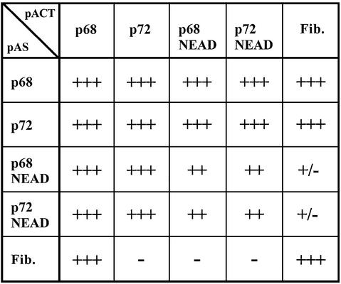
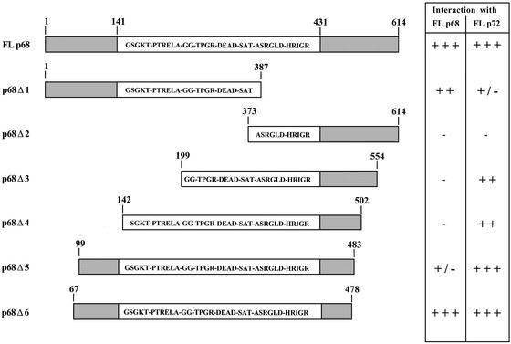
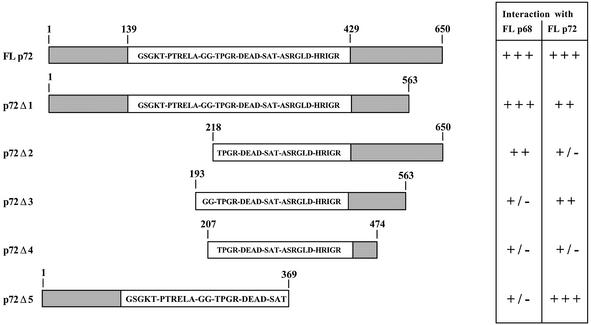
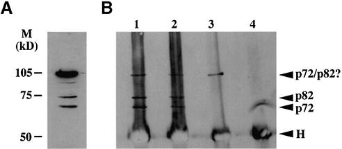
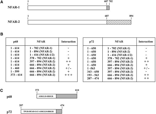
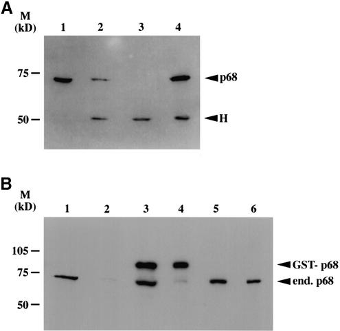
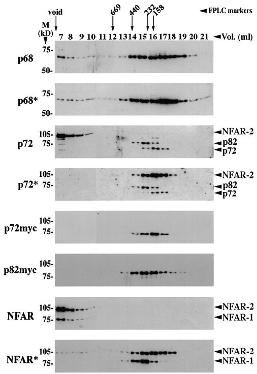
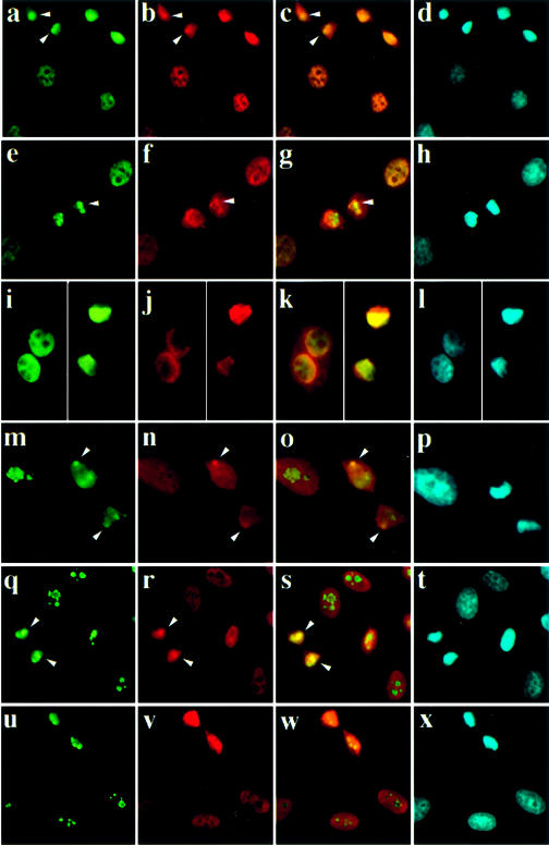
The RNA helicases p68 and p72 are highly related members of the DEAD box family of proteins, sharing 90% identity across the conserved core, and have been shown to be involved in both transcription and mRNA processing. We previously showed that these proteins co-localise in the nucleus of interphase cells. In this study we show that p68 and p72 can interact with each other and self-associate in the yeast two-hybrid system. Co-immunoprecipitation experiments confirmed that p68 and p72 can interact in the cell and indicated that these proteins preferentially exist as hetero-dimers. In addition, we show that p68 can interact with NFAR-2, a protein that is also thought to function in mRNA processing. Moreover, gel filtration analysis suggests that p68 and p72 can exist in a variety of complexes in the cell (ranging from approximately 150 to approximately 400 kDa in size), with a subset of p68 molecules being in very large complexes (>2 MDa). The potential to exist in different complexes that may contain p68 and/or p72, together with a range of other factors, would provide the potential for these proteins to interact with different RNA substrates and would be consistent with recent reports implying a wide range of functions for p68/p72.
Figures
Similar articles
-
The p68 and p72 DEAD box RNA helicases interact with HDAC1 and repress transcription in a promoter-specific manner.BMC Mol Biol. 2004 Aug 6;5:11. doi: 10.1186/1471-2199-5-11. BMC Mol Biol. 2004. PMID: 15298701 Free PMC article.
-
p72: a human nuclear DEAD box protein highly related to p68.Nucleic Acids Res. 1996 Oct 1;24(19):3739-47. doi: 10.1093/nar/24.19.3739. Nucleic Acids Res. 1996. PMID: 8871553 Free PMC article.
-
The nuclear DEAD box RNA helicase p68 interacts with the nucleolar protein fibrillarin and colocalizes specifically in nascent nucleoli during telophase.Exp Cell Res. 2000 Jun 15;257(2):272-80. doi: 10.1006/excr.2000.4886. Exp Cell Res. 2000. PMID: 10837141
-
RNA helicases p68 and p72: multifunctional proteins with important implications for cancer development.Future Oncol. 2011 Feb;7(2):239-51. doi: 10.2217/fon.11.1. Future Oncol. 2011. PMID: 21345143 Review.
-
The DEAD box proteins DDX5 (p68) and DDX17 (p72): multi-tasking transcriptional regulators.Biochim Biophys Acta. 2013 Aug;1829(8):756-63. doi: 10.1016/j.bbagrm.2013.03.004. Epub 2013 Mar 19. Biochim Biophys Acta. 2013. PMID: 23523990 Review.
Cited by
-
The RNA helicase Ddx5/p68 binds to hUpf3 and enhances NMD of Ddx17/p72 and Smg5 mRNA.Nucleic Acids Res. 2013 Sep;41(16):7875-88. doi: 10.1093/nar/gkt538. Epub 2013 Jun 20. Nucleic Acids Res. 2013. PMID: 23788676 Free PMC article.
-
The active form of Xp54 RNA helicase in translational repression is an RNA-mediated oligomer.Nucleic Acids Res. 2004 Feb 24;32(4):1325-34. doi: 10.1093/nar/gkh303. Print 2004. Nucleic Acids Res. 2004. PMID: 14982957 Free PMC article.
-
DEAD-ly Affairs: The Roles of DEAD-Box Proteins on HIV-1 Viral RNA Metabolism.Front Cell Dev Biol. 2022 Jun 13;10:917599. doi: 10.3389/fcell.2022.917599. eCollection 2022. Front Cell Dev Biol. 2022. PMID: 35769258 Free PMC article. Review.
-
RNA Helicase DDX17 Inhibits Hepatitis B Virus Replication by Blocking Viral Pregenomic RNA Encapsidation.J Virol. 2021 Sep 9;95(19):e0044421. doi: 10.1128/JVI.00444-21. Epub 2021 Sep 9. J Virol. 2021. PMID: 34287051 Free PMC article.
-
RNA helicases, DDX5 and DDX17, facilitate lytic reactivation of gammaherpesviruses.PLoS Pathog. 2025 Apr 21;21(4):e1013009. doi: 10.1371/journal.ppat.1013009. eCollection 2025 Apr. PLoS Pathog. 2025. PMID: 40257982 Free PMC article.
References
-
- Linder P., Lasko,P.F., Leroy,P., Nielsen,P.J., Nishi,K., Schnier,J. and Slominski,P.P. (1989) Birth of the D-E-A-D box. Nature, 337, 121–122. - PubMed
-
- Fuller-Pace F.V. (1994) RNA helicases: modulators of RNA structure. Trends Cell Biol., 4, 271–274. - PubMed
-
- de la Cruz J., Kressler,D. and Linder,P. (1999) Unwinding RNA in Saccharomyces cerevisiae: DEAD-box proteins and related families. Trends Biochem. Sci., 24, 192–198. - PubMed
-
- Lane D.P. and Hoeffler,W.K. (1980) SV40 large T shares an antigenic determinant with a cellular protein of molecular weight 68,000. Nature, 288, 167–170. - PubMed
-
- Ford M.J., Anton,I.A. and Lane,D.P. (1988) Nuclear protein with sequence homology to translation initiation factor eIF-4A. Nature, 332, 736–738. - PubMed
Publication types
MeSH terms
Substances
Grants and funding
LinkOut - more resources
Full Text Sources
Other Literature Sources
Molecular Biology Databases
Miscellaneous
