. 2003 Mar 1;31(5):1576-83.
doi: 10.1093/nar/gkg208.

Ageing and telomeres: a study into organ- and gender-specific telomere shortening

Affiliations

Ageing and telomeres: a study into organ- and gender-specific telomere shortening

H Cherif et al. Nucleic Acids Res. .

Abstract

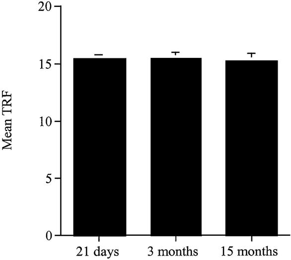
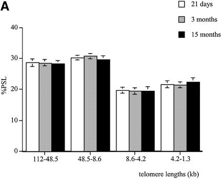
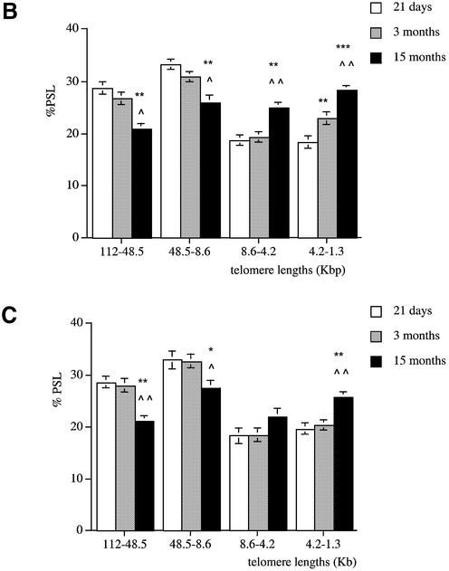
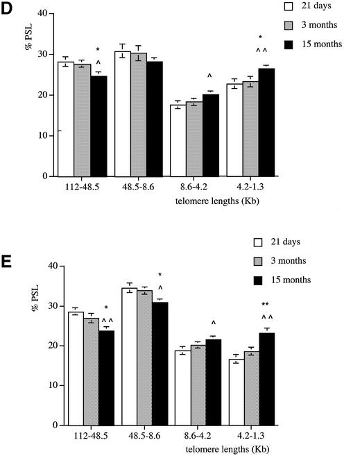
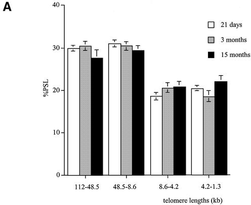
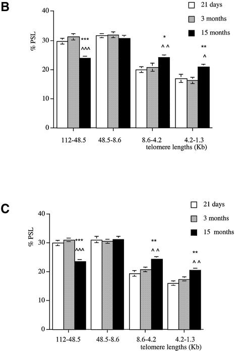
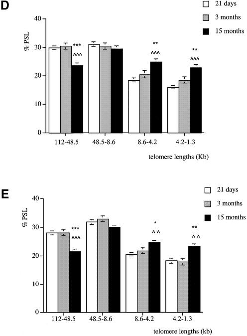
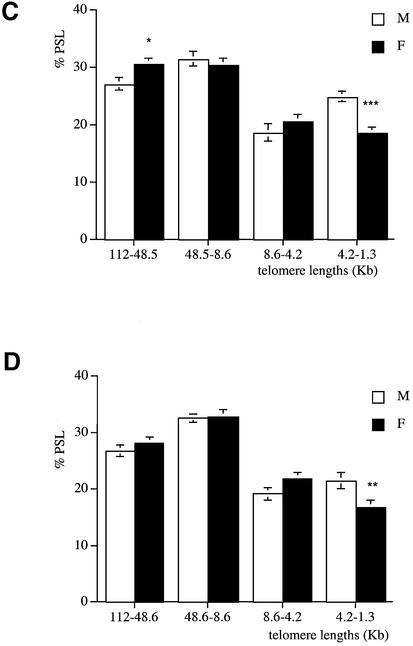
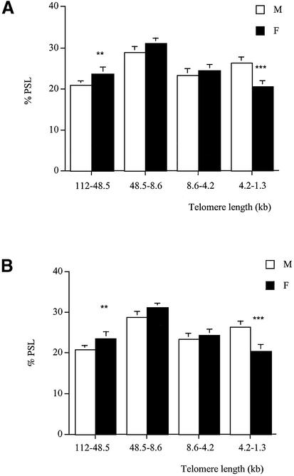
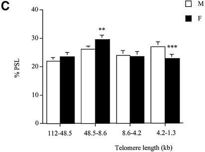
Telomeres, the non-coding sequences at the ends of chromosomes, in the absence of telomerase, progressively shorten with each cell division. Shortening of telomeres can induce cell cycle arrest and apoptosis. The aim of this study was to investigate age- and gender-related changes in telomere length in the rat and to detect possible tissue- specific rates of telomere shortening. Changes with age in telomere lengths were assessed by Southern blotting in the kidney, pancreas, liver, lung and brain of male and female rats. We determined the percentage of telomeres in various molecular size regions rather than measuring the average telomere length. The latter was unable to detect telomere shortening in the tissues. The percentage of short telomeres increased with age in the kidney, liver, pancreas and lung of both males and females, but not in the brain. Males had shorter telomeres than females in all organs analysed except the brain, where the lengths were similar. These findings indicate that telomeres shorten in the rat kidney, liver, pancreas and the lung in an age-dependent manner. These data also provide a novel mechanism for the gender-related differences in lifespan and suggest a tissue-specific regulation of telomere length during development and ageing in the rat.


Figures

Figure 1
Figure 1
Average telomere length (TRF) in the kidney of 21-day-old and 3- and 15-month-old male Wistar rats. Analysis was performed on the same gels used in this study. The TRF in the kidneys was calculated at 21 days and 3 and 15 months.
Figure 2
Figure 2
Telomere shortening with age in male Wistar rats. Genomic DNA was extracted from (A) brain, (B) kidney, (C) liver, (D) pancreas and (E) lung at 21 days (white columns), 3 months (grey columns) and 15 months (black columns). Each telomere smear was divided into four regions according to a molecular weight marker: 112–48.5, 48.5–8.6, 8.6–4.2 and 4.2–1.3 kb. Values are means of n = 8 ± SEM. *P < 0.05, **P < 0.01 and ***P < 0.001 compared to the 21-day-old group; P < 0.05, ∧∧P < 0.01 and ∧∧∧P < 0.001 compared to the 3-month-old group using ANOVA 2 test.
Figure 2
Figure 2
Telomere shortening with age in male Wistar rats. Genomic DNA was extracted from (A) brain, (B) kidney, (C) liver, (D) pancreas and (E) lung at 21 days (white columns), 3 months (grey columns) and 15 months (black columns). Each telomere smear was divided into four regions according to a molecular weight marker: 112–48.5, 48.5–8.6, 8.6–4.2 and 4.2–1.3 kb. Values are means of n = 8 ± SEM. *P < 0.05, **P < 0.01 and ***P < 0.001 compared to the 21-day-old group; P < 0.05, ∧∧P < 0.01 and ∧∧∧P < 0.001 compared to the 3-month-old group using ANOVA 2 test.
Figure 3
Figure 3
Telomere shortening with age in female Wistar rats. Genomic DNA was extracted from (A) brain, (B) kidney, (C) liver, (D) pancreas and (E) lung at 21 days (white columns), 3 months (grey columns) and 15 months (black columns). Each telomere smear was divided into four regions according to a molecular weight marker: 112–48.5, 48.5–8.6, 8.6–4.2 and 4.2–1.3 kb. Values are means of n = 8 ± SEM. *P < 0.05, **P < 0.01 and ***P < 0.001 compared to the 21-day-old group; P < 0.05, ∧∧P < 0.01 and ∧∧∧P < 0.001 compared to the 3-month-old group using ANOVA 2 test.
Figure 3
Figure 3
Telomere shortening with age in female Wistar rats. Genomic DNA was extracted from (A) brain, (B) kidney, (C) liver, (D) pancreas and (E) lung at 21 days (white columns), 3 months (grey columns) and 15 months (black columns). Each telomere smear was divided into four regions according to a molecular weight marker: 112–48.5, 48.5–8.6, 8.6–4.2 and 4.2–1.3 kb. Values are means of n = 8 ± SEM. *P < 0.05, **P < 0.01 and ***P < 0.001 compared to the 21-day-old group; P < 0.05, ∧∧P < 0.01 and ∧∧∧P < 0.001 compared to the 3-month-old group using ANOVA 2 test.
Figure 4
Figure 4
Telomere length analysis reflects gender differences at 3 months of age. Genomic DNA was extracted from (A) kidney, (B) liver, (C) pancreas and (D) lung of males (white columns) and females (black columns). Each telomere smear was divided into four regions according to a molecular weight marker: 112–48.5, 48.5–8.6, 8.6–4.2 and 4.2–1.3 kb. Arbitrary units (% PSL) were calculated for each size region. Values are means of n = 8 ± SEM. *P < 0.05, **P < 0.01 and ***P < 0.001 using the unpaired Student’s t-test.
Figure 4
Figure 4
Telomere length analysis reflects gender differences at 3 months of age. Genomic DNA was extracted from (A) kidney, (B) liver, (C) pancreas and (D) lung of males (white columns) and females (black columns). Each telomere smear was divided into four regions according to a molecular weight marker: 112–48.5, 48.5–8.6, 8.6–4.2 and 4.2–1.3 kb. Arbitrary units (% PSL) were calculated for each size region. Values are means of n = 8 ± SEM. *P < 0.05, **P < 0.01 and ***P < 0.001 using the unpaired Student’s t-test.
Figure 5
Figure 5
Telomere length analysis reflects gender differences at 15 months of age. Genomic DNA was extracted from (A) kidney, (B) liver and (C) pancreas of males (white columns) and females (black columns). Each telomere smear was divided into four regions according to a molecular weight marker: 112–48.5, 48.5–8.6, 8.6–4.2 and 4.2–1.3 kb. Arbitrary units (% PSL) were calculated for each size region. Values are means of n = 8 ± SEM. **P < 0.01 and ***P < 0.001 using the unpaired Student’s t-test.
Figure 5
Figure 5
Telomere length analysis reflects gender differences at 15 months of age. Genomic DNA was extracted from (A) kidney, (B) liver and (C) pancreas of males (white columns) and females (black columns). Each telomere smear was divided into four regions according to a molecular weight marker: 112–48.5, 48.5–8.6, 8.6–4.2 and 4.2–1.3 kb. Arbitrary units (% PSL) were calculated for each size region. Values are means of n = 8 ± SEM. **P < 0.01 and ***P < 0.001 using the unpaired Student’s t-test.

References

    1. Meyne J., Ratliff,R.L. and Moysis,R.K. (1989) Conservation of the human telomere sequence (TTAGGG)n among vertebrates. Proc. Natl Acad. Sci. USA, 86, 7049–7053. - PMC - PubMed
    1. Zakian V.A. (1996) Structure, function and replication of Saccharomyces cerevisiae telomeres. Annu. Rev. Genet., 30, 141–172. - PubMed
    1. Greider C.W. (1994) Identification of a specific telomere terminal transferase activity in Tetrahymena extracts. Curr. Opin. Genet. Dev., 4, 203–211. - PubMed
    1. Greider C.W. and Blackburn,E.H. (1985) Mammalian telomere dynamics: healing, fragmentation, shortening and stabilisation. Cell, 43, 405–413. - PubMed
    1. Yu G.-L., Bradley,J.D., Attardi,L.D. and Blackburn,E.H. (1990) In vivo alteration of telomere sequences and senescence caused by mutated Tetrahymena telomerase RNAs. Nature, 344, 126–132. - PubMed

Publication types