. 2003 Mar 1;31(5):e22.
doi: 10.1093/nar/gng022.

New restriction enzymes discovered from Escherichia coli clinical strains using a plasmid transformation method

Affiliations

New restriction enzymes discovered from Escherichia coli clinical strains using a plasmid transformation method

Julie K A Kasarjian et al. Nucleic Acids Res. .

Abstract

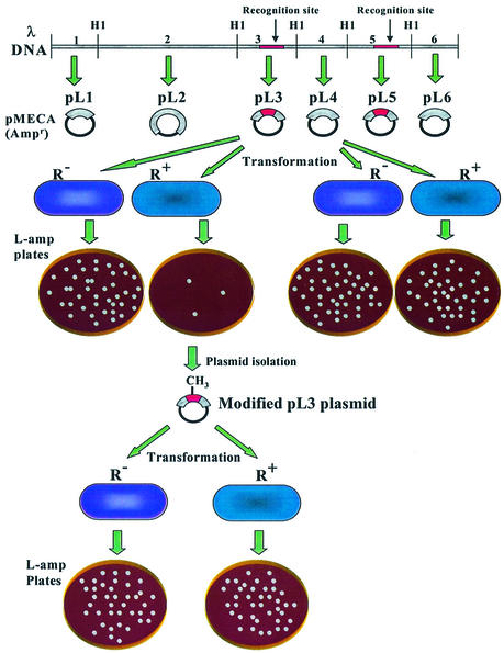
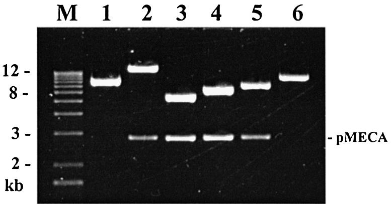
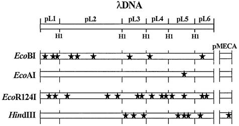
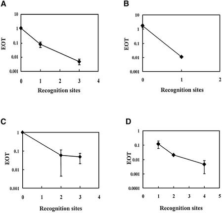
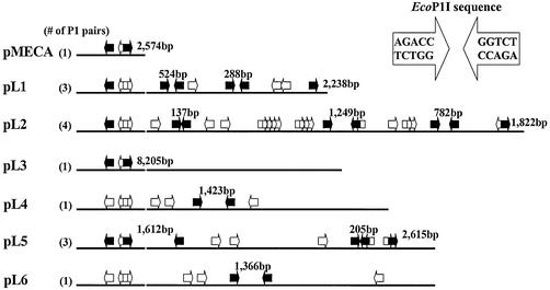
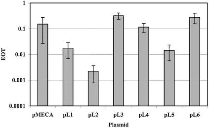
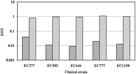
The presence of restriction enzymes in bacterial cells has been predicted by either classical phage restriction-modification (R-M) tests, direct in vitro enzyme assays or more recently from bacterial genome sequence analysis. We have applied phage R-M test principles to the transformation of plasmid DNA and established a plasmid R-M test. To validate this test, six plasmids that contain BamHI fragments of phage lambda DNA were constructed and transformed into Escherichia coli strains containing known R-M systems including: type I (EcoBI, EcoAI, Eco124I), type II (HindIII) and type III (EcoP1I). Plasmid DNA with a single recognition site showed a reduction of relative efficiency of transformation (EOT = 10(-1)-10(-2)). When multiple recognition sites were present, greater reductions in EOT values were observed. Once established in the cell, the plasmids were subjected to modification (EOT = 1.0). We applied this test to screen E.coli clinical strains and detected the presence of restriction enzymes in 93% (14/15) of cells. Using additional subclones and the computer program, RM Search, we identified four new restriction enzymes, Eco377I, Eco585I, Eco646I and Eco777I, along with their recognition sequences, GGA(8N)ATGC, GCC(6N)TGCG, CCA(7N)CTTC, and GGA(6N)TATC, respectively. Eco1158I, an isoschizomer of EcoBI, was also found in this study.


Figures

Figure 1
Figure 1
A diagram showing the subcloning of the six lambda BamHI (HI) fragments into pMECA (top) and the plasmid restriction (center) and modification test (bottom) using a transformation method. In this figure, it is assumed that lambda BamHI fragments no. 3 (cloned in pL3) and no. 5 (pL5) each contain a recognition site (darkened area), whereas fragment no. 4 (pL4) and the remaining fragments do not contain a restriction site. Plasmid pMECA is a cloning vector and is represented as a solid line. R+ bacteria produce a restriction enzyme that cleaves pL3, which has an unmodified recognition site. R bacteria have no restriction activity and are used as a control for the restriction test. Modified pL3 can be isolated from surviving colonies following transformation. Using this modified pL3, a modification test can be performed by transforming this plasmid to both R and R+ strains. Modified plasmids are expected to result in the same number of transformants in both strains.
Figure 2
Figure 2
pL1 to pL6 plasmid subclones derived from phage lambda. To construct the pL series of subclones, lambda DNA was digested with BamHI and cloned into the BamHI site of pMECA. The subclones are shown here after digestion with BamHI. Lane M, 1 kb ladder; lanes 1–6, pL1–pL6. Lanes 2–5 show both the inserts (16.8, 5.6, 6.5, 7.2 kb, respectively) and the pMECA vector (2.8 kb). Plasmids pL1 and pL6 (5.8 and 7.2 kb inserts, respectively) contain a single BamHI site and include the end clones of phage lambda.
Figure 3
Figure 3
Locations of type I and type II recognition sequences in pMECA and lambda DNA. Each black star represents one recognition site. Plasmids pL1–pL6 each contain a BamHI fragment of lambda DNA. Note that the vector, pMECA, contains one HindIII site. Map not to scale.
Figure 4
Figure 4
Relationship between EOT and the number of recognition sequences present in plasmid DNA. Average EOT values are plotted against the number of recognition sequences for R-M systems EcoBI (A), EcoAI (B), EcoR124I (C) and HindIII (D). Average EOT values and standard deviations were calculated from the data shown in Table 1.
Figure 5
Figure 5
Possible EcoP1I sites present in pMECA and the pL series plasmids. Plasmid DNA is shown in a linear form after digestion with BamHI. Type III R-M systems require a pair of inversely oriented EcoP1I sites. Each arrow represents one EcoP1I site. A P1 pair, formed of two inversely oriented sites, is shown in the inset. Pairs separated by a distance of <3.5 kb are shown as solid arrows. In some cases, additional pairs can be formed using a similar end. For clarity, only the shortest pairs are counted in the parentheses at the left.
Figure 6
Figure 6
EcoP1I plasmid restriction test. All plasmids were transformed into strain WA921 using E.coli C as a control. Modified plasmids were then obtained and transformed into each strain. Details of the EOT calculation are described in the Materials and Methods.
Figure 7
Figure 7
Recognition sequence confirmation using plasmid R-M test. EOT data for R-M tests are shown as solid and striped bars, respectively. Bacterial strain DH5α and plasmid pMECA were used as controls.

Similar articles

Cited by

References

    1. Arber W. and Linn,S. (1969) DNA modification and restriction. Annu. Rev. Biochem., 38, 467–500. - PubMed
    1. Murray N.E. (2000) Type I restriction systems: sophisticated molecular machines (a legacy of Bertani and Weigle). Microbiol. Mol. Biol. Rev., 64, 412–434. - PMC - PubMed
    1. Redaschi N. and Bickle,T.A. (1996) DNA restriction and modification systems. In Neidhardt,F.C. (ed.), Escherichia coli and Salmonella typhimurium: Cellular and Molecular Biology. ASM Press, Washington, DC, pp. 773–781.
    1. Roberts R.J. and Macelis,D. (2001) REBASE—restriction enzymes and methylases. Nucleic Acids Res., 29, 268–269. - PMC - PubMed
    1. Kong H., Lin,L.F., Porter,N., Stickel,S., Byrd,D., Posfai,J. and Roberts,R.J. (2000) Functional analysis of putative restriction-modification system genes in the Helicobacter pylori J99 genome. Nucleic Acids Res., 28, 3216–3223. - PMC - PubMed

Publication types

MeSH terms

Substances