Studies on the molecular pathogenesis of extraskeletal myxoid chondrosarcoma-cytogenetic, molecular genetic, and cDNA microarray analyses
- PMID: 12598313
- PMCID: PMC1868116
- DOI: 10.1016/S0002-9440(10)63875-8
Studies on the molecular pathogenesis of extraskeletal myxoid chondrosarcoma-cytogenetic, molecular genetic, and cDNA microarray analyses
Abstract
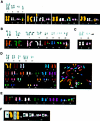
Extraskeletal myxoid chondrosarcomas (EMCs) are characterized by recurrent chromosome translocations resulting in fusions of the nuclear receptor TEC to various NH(2)-terminal partners. Here we describe the phenotypic, cytogenetic, and molecular genetic characteristics of a series of 10 EMCs. Using spectral karyotyping and fluorescence in situ hybridization, clonal chromosome abnormalities were detected in all but one tumor. A t(9;22)(q22;q12) translocation was found in three cases; a del(22)(q12-13)in one case; and variant translocations, including t(9;17)(q22;q11-12), t(7;9;17)(q32;q22;q11), and t(9;15)(q22;q21), were detected in one case each. Recurrent, secondary abnormalities, including trisomy 1q, 7, 8, 12, and 19, were found in seven tumors. All tumors contained translocation-generated or cryptic gene fusions, including EWS-TEC (five cases, of which one was a novel fusion), TAF2N-TEC (four cases), and TCF12-TEC (one case). cDNA microarray analysis of the gene expression patterns of two EMCs and a myxoid liposarcoma reference tumor revealed a remarkably distinct and uniform expression profile in both EMCs despite the fact that they had different histologies and expressed different fusion transcripts. The most differentially expressed gene in both tumors was CHI3L1, which encodes a secreted glycoprotein (YKL-40) previously implicated in various pathological conditions of extracellular matrix degradation as well as in cancer. Our findings suggests that EMC exhibits a tumor-specific gene expression profile, including overexpression of several cancer-related genes as well as genes implicated in chondrogenesis and neural-neuroendocrine differentiation, thus distinguishing it from other soft tissue sarcomas.
Figures


Similar articles
-
Fusion of the NH2-terminal domain of the basic helix-loop-helix protein TCF12 to TEC in extraskeletal myxoid chondrosarcoma with translocation t(9;15)(q22;q21).Cancer Res. 2000 Dec 15;60(24):6832-5. Cancer Res. 2000. PMID: 11156374
-
Fusion of the EWS-related gene TAF2N to TEC in extraskeletal myxoid chondrosarcoma.Cancer Res. 1999 Oct 15;59(20):5064-7. Cancer Res. 1999. PMID: 10537274
-
Extraskeletal myxoid chondrosarcoma: multimodal diagnosis and identification of a new cytogenetic subgroup characterized by t(9;17)(q22;q11).Virchows Arch. 1999 Nov;435(5):524-30. doi: 10.1007/s004280050437. Virchows Arch. 1999. PMID: 10592057
-
t(9;22)(q22-31;q11-12) is a consistent marker of extraskeletal myxoid chondrosarcoma: evaluation of three cases.Mod Pathol. 1995 Sep;8(7):765-8. Mod Pathol. 1995. PMID: 8539235 Review.
-
Genetics of chondrosarcoma and related tumors.Curr Opin Oncol. 2004 Jul;16(4):342-54. doi: 10.1097/01.cco.0000129678.72521.e5. Curr Opin Oncol. 2004. PMID: 15187889 Review.
Cited by
-
Differential expression of Chitinase 3-Like 1 protein in appendicitis and appendix carcinomas.J Clin Lab Anal. 2022 Dec;36(12):e24790. doi: 10.1002/jcla.24790. Epub 2022 Nov 29. J Clin Lab Anal. 2022. PMID: 36447423 Free PMC article.
-
Curious case of extraskeletal myxoid chondrosarcoma.Lung India. 2017 Mar-Apr;34(2):170-172. doi: 10.4103/0970-2113.201312. Lung India. 2017. PMID: 28360467 Free PMC article.
-
Cytogenetics and molecular genetics of myxoid soft-tissue sarcomas.Genet Res Int. 2011;2011:497148. doi: 10.4061/2011/497148. Epub 2011 Jul 28. Genet Res Int. 2011. PMID: 22567356 Free PMC article.
-
Extraskeletal myxoid chondrosarcoma with small bowel metastasis causing bowel obstruction.Case Rep Oncol Med. 2012;2012:621025. doi: 10.1155/2012/621025. Epub 2012 Nov 18. Case Rep Oncol Med. 2012. PMID: 23213584 Free PMC article.
-
Top Ten Differentials to Mull Over for Head and Neck Myoepithelial Neoplasms.Head Neck Pathol. 2023 Mar;17(1):1-15. doi: 10.1007/s12105-022-01502-0. Epub 2023 Mar 16. Head Neck Pathol. 2023. PMID: 36928733 Free PMC article. Review.
References
-
- Enzinger FM, Shiraki M: Extraskeletal myxoid chondrosarcoma: an analysis of 34 cases. Hum Pathol 1972, 3:421-435 - PubMed
-
- Meis-Kindblom JM, Bergh P, Gunterberg B, Kindblom L-G: Extraskeletal myxoid chondrosarcoma: a reappraisal of its morphologic spectrum and prognostic factors based on 117 cases. Am J Surg Pathol 1999, 23:636-650 - PubMed
-
- Kindblom LG, Angervall L: Histochemical characterization of mucosubstances in bone and soft tissue-tumors. Cancer 1975, 36:985-994 - PubMed
-
- Harris M, Coyne J, Tariq M, Eyden BP, Atkinson M, Freemont AJ, Varley J, Attwooll C, Telford N: Extraskeletal myxoid chondrosarcoma with neuroendocrine differentiation: a pathologic, cytogenetic, and molecular study of a case with a novel translocation t(9;17)(q22;q11.2). Am J Surg Pathol 2000, 24:1020-1026 - PubMed
-
- Okamoto S, Hisaoka M, Ishida T, Imamura T, Kanda H, Shimajiri S, Hashimoto H: Extraskeletal myxoid chondrosarcoma: a clinicopathologic, immunohistochemical, and molecular analysis of 18 cases. Hum Pathol 2001, 32:1116-1124 - PubMed
Publication types
MeSH terms
LinkOut - more resources
Full Text Sources
Other Literature Sources

