. 2003 Mar;162(3):837-47.
doi: 10.1016/S0002-9440(10)63880-1.

AIM inhibits apoptosis of T cells and NKT cells in Corynebacterium-induced granuloma formation in mice

Affiliations

AIM inhibits apoptosis of T cells and NKT cells in Corynebacterium-induced granuloma formation in mice

Kazuhisa Kuwata et al. Am J Pathol. 2003 Mar.

Abstract

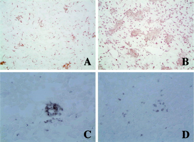
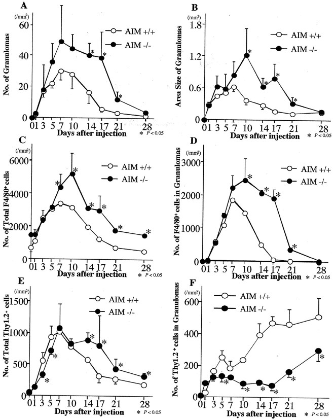
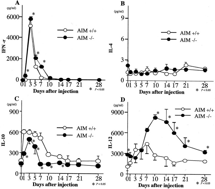
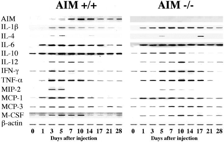
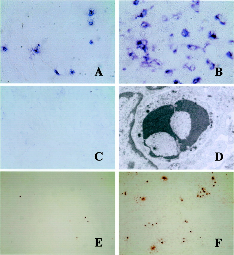
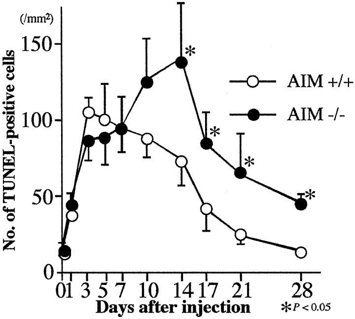
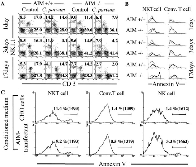
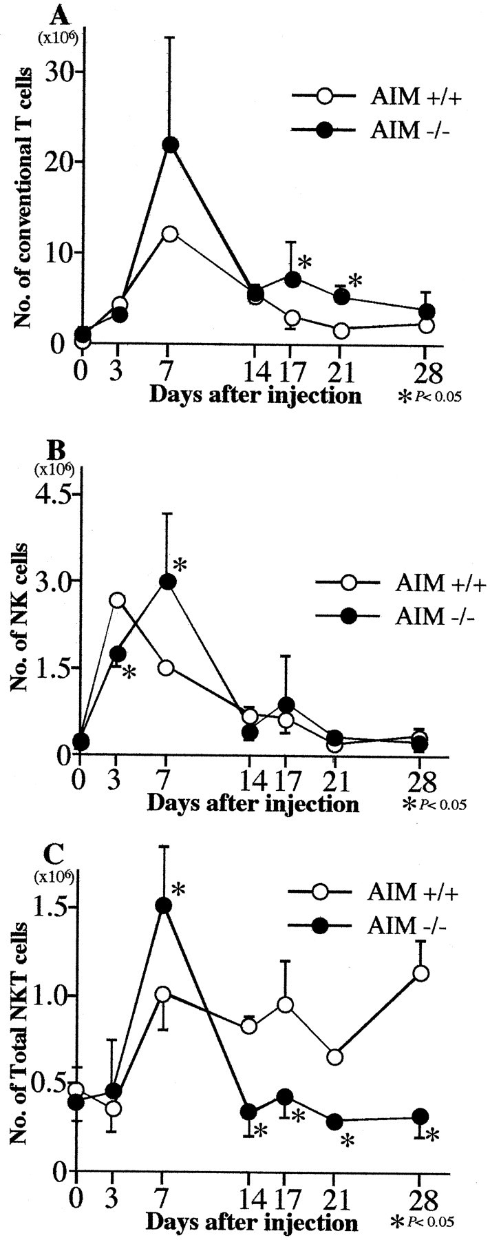
Apoptosis inhibitor expressed by macrophages (AIM) inhibits apoptosis of CD4(+)CD8(+) (CD4/CD8) double-positive thymocytes, and supports the viability of these cells on the thymic selection. However, pleiotropic functions of AIM have been suggested. In this study, heat-killed Corynebacterium parvum (C. parvum) was injected into mice carrying the homozygous mutation (AIM(-/-)) and wild-type (AIM(+/+)) mice, to investigate the role of AIM in the formation of hepatic granulomas. In AIM(-/-) mice, the size and the number of hepatic granulomas were larger, and the resorption of granulomas was more delayed than in AIM(+/+) mice. The production of interleukin-12 was more prominent in AIM(-/-) mice than in AIM(+/+) mice. In the liver of AIM(+/+) mice, expression of AIM messenger ribonucleic acid (mRNA) increased after C. parvum injection. In situ hybridization demonstrated that AIM mRNA was expressed in Kupffer cells and exudate macrophages in the liver, especially in granulomas. Larger numbers of T cells and natural killer T (NKT) cells underwent apoptosis in the granulomas of AIM(-/-) mice, suggesting that AIM prevents apoptosis of NKT cells and T cells in C. parvum-induced inflammation. Recombinant AIM (rAIM) protein significantly inhibited apoptosis of NKT cells and T cells obtained from C. parvum-stimulated livers in vitro. These results indicate that AIM functions to induce resistance to apoptosis within NKT cells and T cells, and supports the host defense in granulomatous inflammation.


Figures

Figure 1.
Figure 1.
Immunohistochemistry of C. parvum-injected livers of homozygous mutant (AIM−/−) (B and D) and wild-type (AIM+/+) mice (A and C) at 17 days after injection. A and B: Accumulations of F4/80+ macrophages and granuloma formation are more prominent in an AIM−/− mouse (B) than in an AIM+/+ mouse (A). Original magnification, ×100. C and D: Infiltration of Thy1.2+ T cells in the granulomas of an AIM−/− mouse (D) is less remarkable than that of an AIM+/+ mouse (C). Original magnification, ×200.
Figure 2.
Figure 2.
The number (A) and area size (B) of granulomas in livers, the numbers of macrophages in the liver (C) and in the granulomas (D), and the numbers of Thy 1.2+ T cells in the liver (E) and in the granulomas (F) of AIM−/− and AIM+/+ mice after C. parvum injection. A and B: AIM−/− mice develop larger numbers of granulomas than AIM+/+ mice. C and D: Larger numbers of macrophages were present in the liver and granulomas in AIM−/− mice than AIM+/+ mice. E and F: The numbers of T cells had not remarkable difference in the liver, but there were smaller numbers of T cells in the granulomas of AIM−/− than in AIM+/+ mice. Data are shown as the mean ± SD of five mice. *, P < 0.05.
Figure 3.
Figure 3.
Serum concentrations of IFN-γ (A), IL-4 (B), IL-10 (C), and IL-12 (D) in AIM−/− and AIM+/+ mice after C. parvum injection. Serum levels of IL-12 and IFN-γ were higher in AIM−/− than in AIM+/+ mice. Serum levels of IL-10 were lower in AIM−/− than AIM+/+ mice. Data are shown as the mean ± SD of three mice. *, P < 0.05.
Figure 4.
Figure 4.
Expression of AIM, cytokines, and β-actine mRNAs of AIM−/− and AIM+/+ mice after C. parvum injection. The result from one of three similar experiments is shown.
Figure 5.
Figure 5.
AIM mRNA expression in the liver by in situ hybridization (A-C), electron micrograph of an apoptotic lymphocyte (D), and TUNEL-staining of apoptotic cells (E and F). A–C: AIM mRNA is weakly expressed in a few round or spindle cells along the hepatic sinusoids of untreated AIM+/+ mice (A). At day 17 after C. parvum injection, AIM expression is augmented in macrophages (B), especially in granulomas. However, signal-positive cells were not detected in the liver of C. parvum-injected and non-injected AIM−/− mice (C). Original magnification, ×400. D: Electron micrograph of a lymphocyte in the granuloma with small electron-dense intracytoplasmic granules. Condensed nucleus suggests apoptosis of this granular lymphocyte. Original magnification, ×12,000. E and F: At 17 days TUNEL-labeled cells are more abundant in the liver of AIM−/− mice (F) than in AIM+/+ mice (E). Original magnification, ×40.
Figure 6.
Figure 6.
The numbers of TUNEL-labeled cells in the liver of AIM−/− and AIM+/+ mice after C. parvum injection. After 7 days, larger numbers of apoptotic cells were present in the liver of AIM−/− mice than AIM+/+ mice. Data are shown as the mean ± SD of three mice. *, P < 0.05.
Figure 7.
Figure 7.
Phenotypic characterization of T cell in the liver of AIM−/− and AIM+/+ mice by flow cytometric analysis for CD3 and NK1.1 (A) and for CD3, NK1.1, and Annexin V expression (B) at 1, 3, and 17 days after C. parvum injection. Surface phenotype analysis of cultured cells; liver MNCs (1 × 10⁷ cells per well) were cultured in 24-well microculture plates with (lower) or without (upper) rAIM protein for 24 hours (C). A: Proportion of NKT cells were decreased in both mice after C. parvum injection. However, repopulation of NKT cells was not observed in the knockout mice at 17 days. Numbers indicate the percentage of fluorescence-positive cells in the corresponding squares. The right upper gates exhibit the population of NKT cells, and the right lower gates show these of conventional T cells. Typical results from two experiments are shown. B: Apoptotic elimination of hepatic NKT cells and conventional T cells in the liver of AIM−/− was more remarkable than that of AIM+/+ mice, especially at 1 day and 17 days after C. parvum injection. Annexin V-positive NKT cells and conventional T cells in the liver of both mice after C. parvum injection are represented by bold histograms, and Annexin V-positive NKT cells and conventional T cells in the liver of each untreated mice are represented by thin histograms. Annexin V-positive cells are induced apoptosis. C: Cultured NKT cells with rAIM protein were more diminished in the number and proportion of apoptotic cells, and a few conventional T cells, but not NK cells. Percentages of labeled cells with Annexin V are shown in the histogram. Mean fluorescence intensities are shown in the brackets.
Figure 8.
Figure 8.
The absolute cell numbers of conventional T cells (A), NK cells (B), and NKT cells (C) in the liver of AIM−/− and AIM+/+ mice after C. parvum injection. The numbers of NKT cells in the liver of AIM−/− mice became smaller than AIM+/+ mice from 14 days after C. parvum injection. The absolute cell numbers were calculated on total liver MNCs and the cell subsets obtained from flow cytometric analysis with anti-CD3 and anti-NK1.1 staining. Data are shown as the mean ± SD of three mice. *, P < 0.05.

Similar articles

Cited by

References

    1. Miyazaki T, Hirokami Y, Matsuhashi N, Takatsuka H, Naito M: Increased susceptibility of thymocytes to apoptosis in mice lacking AIM, a novel murine macrophage-derived soluble factor belonging to the scavenger receptor cysteine-rich domain superfamily. J Exp Med 1999, 189:413-422 - PMC - PubMed
    1. Oyama H, Yamada T, Inoue Y: Effect of post-irradiation temperature on viability of rat thymocytes. Int J Radiat Biol Relat Stud Phys Chem Med 1974, 26:535-546 - PubMed
    1. Ohyama H, Yamada T, Ohkawa A, Watanabe I: Radiation-induced formation of apoptotic bodies in rat thymus. Radiat Res 1985, 101:123-130 - PubMed
    1. Bodey B, Bodey B, Jr, Kaiser HE: Apoptosis in the mammalian thymus during normal histogenesis and under various in vitro and in vivo experimental conditions. In Vivo 1998, 12:123-133 - PubMed
    1. Selye H: Thymus and adrenals in the response of the organism to injuries and intoxication. Br J Exp Pathol 1936, 17:234-248

Publication types

MeSH terms