. 2003 Mar;162(3):897-905.
doi: 10.1016/S0002-9440(10)63885-0.

PTOV-1, a novel protein overexpressed in prostate cancer, shuttles between the cytoplasm and the nucleus and promotes entry into the S phase of the cell division cycle

Affiliations

PTOV-1, a novel protein overexpressed in prostate cancer, shuttles between the cytoplasm and the nucleus and promotes entry into the S phase of the cell division cycle

Anna Santamaría et al. Am J Pathol. 2003 Mar.

Abstract

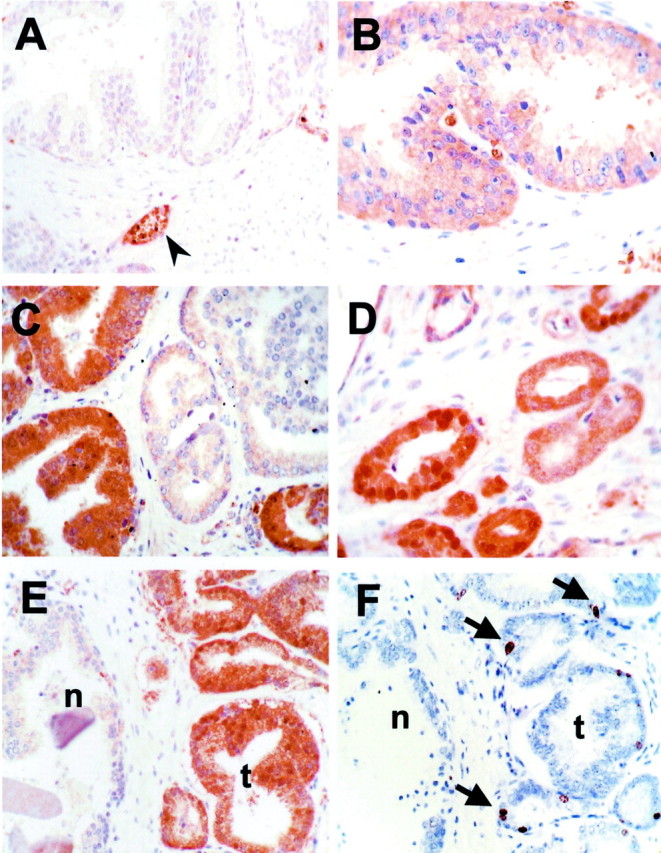
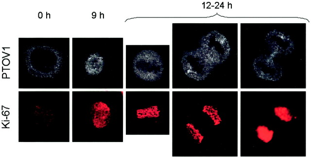
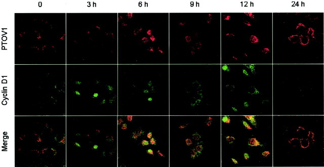
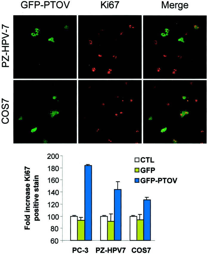
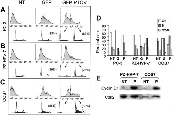
PTOV1 was recently identified as a novel gene and protein during a differential display screening for genes overexpressed in prostate cancer. The PTOV1 protein consists of two novel protein domains arranged in tandem, without significant similarities to known protein motifs. By immunohistochemical analysis, we have found that PTOV1 is overexpressed in 71% of 38 prostate carcinomas and in 80% of samples with prostate intraepithelial neoplasia. High levels of PTOV1 in tumors correlated significantly with proliferative index, as assessed by Ki67 immunoreactivity, and associated with a nuclear localization of the protein, suggesting a functional relationship between PTOV1 overexpression, proliferative status, and nuclear localization. In quiescent cultured prostate tumor cells, PTOV1 localized to the cytoplasm, being excluded from nuclei. After serum stimulation, PTOV1 partially translocated to the nucleus at the beginning of the S phase. At the end of mitosis, PTOV1 exited the nucleus. Transient transfection of chimeric green fluorescent protein-PTOV1 forced the entry of cells into the S phase of the cell cycle, as shown by double fluorescent imaging for green fluorescent protein and for Ki67, and also by flow cytometry. This was accompanied by greatly increased levels of cyclin D1 protein in the transfected cells. These observations suggest that overexpression of PTOV1 can contribute to the proliferative status of prostate tumor cells and thus to their biological behavior.


Figures

Figure 1.
Figure 1.
Immunohistochemical analysis of expression of PTOV1 and Ki67 in normal and tumor samples. A: Staining for PTOV1 of normal prostate epithelium, showing no detectable staining of glands, and a strong staining of blood vessels and red blood cells (arrowhead). B: High-grade PIN, showing a diffuse cytoplasmic staining for PTOV1 (level 2, see text and Materials and Methods). C: Staining for PTOV1 of a tumor sample, showing heterogeneous staining, with a strong positivity (level 3) in some glands, and faint to no staining in other tumoral glands. D: A tumor sample showing strong nuclear and cytoplasmic staining for PTOV1. E and F: Near-serial sections of a tumor sample containing glands with normal (n) and carcinomatous (t) morphology, stained for PTOV1 (E) and for Ki67 (F). Arrows indicate nuclei positive for Ki67 staining. Original magnifications, ×400.
Figure 2.
Figure 2.
Nuclear localization of PTOV1 in proliferating cells. PZ-HPV-7 cells were growth factor-deprived for 48 hours, stimulated with 10% fetal bovine serum for the indicated times, and stained for PTOV1 or Ki67. Growth factor-deprived cells (time 0) that did not show staining for Ki67 (quiescence) showed a cytoplasmic staining for PTOV1 with nuclear exclusion. Nine hours after serum stimulation, coincident with a nuclear staining for Ki67, PTOV1 translocated into the nucleus. In dividing cells (12 hours after stimulation), PTOV1 is excluded from Ki67-stained condensed chromosomes.
Figure 3.
Figure 3.
Nuclear entry of PTOV1 is concomitant with nuclear exit of cyclin D1. PC-3 cells were growth factor-deprived for 48 hours, stimulated with 10% fetal bovine serum for the indicated times, and stained by double fluorescence for PTOV1 and cyclin D1. In quiescent cells (time 0), PTOV1 is localized exclusively in the cytoplasm, whereas cyclin D1 is barely detectable. From 3 to 6 hours after stimulation with serum, cyclin D1 is detectable in the nucleus of most cells. PTOV1 is found in the nucleus at increasing proportion at 9 to 12 hours after serum stimulation, and its nuclear localization is most evident in cells in which cyclin D1 has been excluded from the nucleus (12 hours). At 24 hours after stimulation, cells show either a cytoplasmic or a nuclear localization of PTOV1, together with low levels of cyclin D1.
Figure 4.
Figure 4.
Overexpression of GFP-PTOV1 induces cell proliferation. PZ-HPV-7 and COS7 cells were transfected with GFP-PTOV1 (green fluorescence) and stained for Ki67 (red fluorescence). Cells positive for GFP-PTOV1, Ki67, or both were quantitated, and the relative numbers were compared to untransfected cells (histogram). As controls, cells were transfected with vector alone (GFP).
Figure 5.
Figure 5.
Overexpression of GFP-PTOV1 forces entry into the S phase of the cell cycle. A—C Flow cytometry analysis of untransfected cells (NT), cells transfected with vector alone (GFP), or with GFP-PTOV1. The threshold for positivity for GFP was established as the levels corresponding to untransfected cells. Cells with green fluorescence below (not transfected) or above (transfected) this threshold were analyzed separately for DNA content. The histograms at the top of each panel correspond to GFP levels, and the histograms at the bottom of each panel correspond to analysis of DNA content and cell-cycle assignments. For each experiment, transfection efficiency is shown in brackets. D: Histograms of distribution of cell populations to the G1, S, or G2 to M phases of the cell cycle, corresponding to the flow cytometry analysis on the left panels. One representative experiment of four performed with each cell line is shown. E: Levels of cyclin D1 are elevated in cells transfected with GFP-PTOV1. PZ-HPV-7 or COS7 cells were either not transfected (NT) or transfected (P) with GFP-PTOV1, lysed 24 hours after transfection, and analyzed by Western blotting with antibodies to cyclin D1 or CDK2.

References

    1. Jenster G: The role of the androgen receptor in the development and progression of prostate cancer. Semin Oncol 1999, 26:407-421 - PubMed
    1. Grossmann ME, Huang H, Tindall DJ: Androgen receptor signaling in androgen-refractory prostate cancer. J Natl Cancer Inst 2001, 93:1687-1697 - PubMed
    1. Marcelli M, Ittmann M, Mariani S, Sutherland R, Nigam R, Murthy L, Zhao Y, DiConcini D, Puxeddu E, Esen A, Eastham J, Weigel NL, Lamb DJ: Androgen receptor mutations in prostate cancer. Cancer Res 2000, 60:944-949 - PubMed
    1. Linja MJ, Savinainen KJ, Saramaki OR, Tammela TL, Vessella RL, Visakorpi T: Amplification and overexpression of androgen receptor gene in hormone-refractory prostate cancer. Cancer Res 2001, 61:3550-3555 - PubMed
    1. Taplin ME, Bubley GJ, Shuster TD, Frantz ME, Spooner AE, Ogata GK, Keer HN, Balk SP: Mutation of the androgen-receptor gene in metastatic androgen-independent prostate cancer. N Engl J Med 1995, 332:1393-1398 - PubMed

Publication types

MeSH terms