

Spinal amino acid release and repeated withdrawal in spinal morphine tolerant rats
- PMID: 12598423
- PMCID: PMC1573708
- DOI: 10.1038/sj.bjp.0705102
Spinal amino acid release and repeated withdrawal in spinal morphine tolerant rats
Abstract
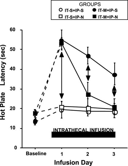
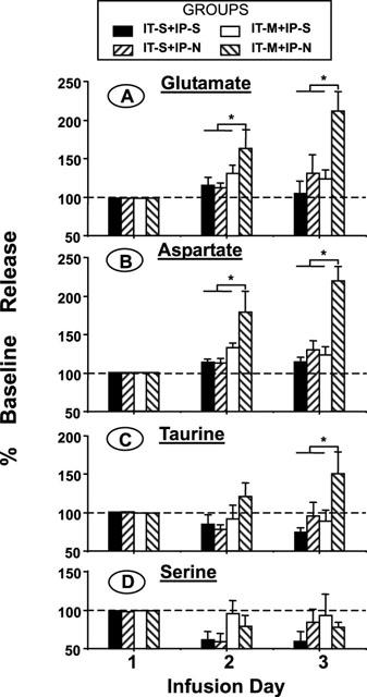
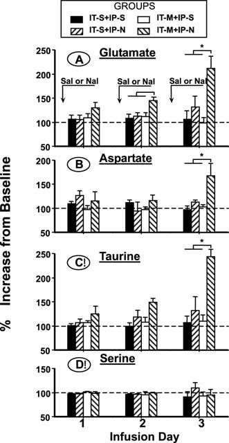
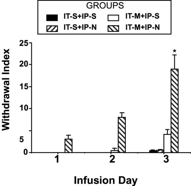
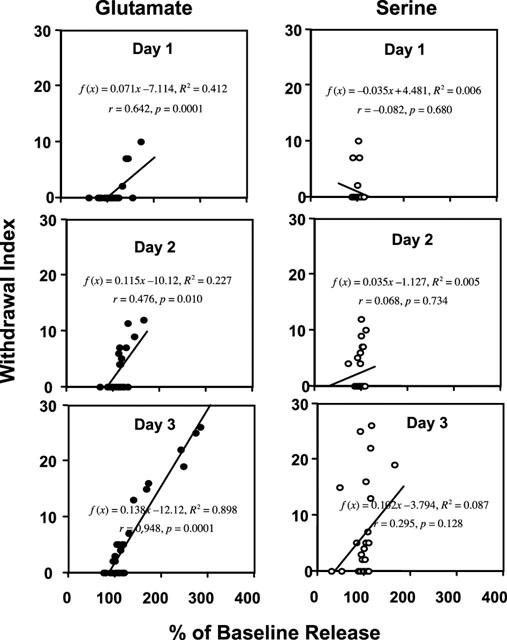
1. We used spinal microdialysis in awake rats to investigate whether the repeated withdrawal with naloxone during continuous spinal infusion of morphine would lead to a progressively greater spinal glutamate release and a more pronounced intrathecal tolerance. 2. Rats received lumbar intrathecal (IT) infusion of morphine (IT-M: 20 nmol microl(-1) h(-1)) or saline (IT-S: 1 microl h(-1)) continuously for 3 days. Both groups were further subdivided to receive intraperitoneal (i.p.) injection of naloxone (IP-N: 0.6 mg kg(-1)) or saline (IP-S: 3 ml kg(-1)) every 24 h after the beginning of IT infusion. Daily thermal escape latencies, withdrawal signs, the resting basal release of spinal amino acids before IP injection and the release immediately after the injection (evoked) were measured. 3. Rats receiving IT morphine showed a maximum increase in thermal escape latency on day 1, after which this value declined, with the fastest decline observed in IT morphine + IP naloxone group. On day 1, no significant difference was observed among groups in the resting basal release of amino acids. Rats in IT morphine + i.p. naloxone group displayed a progressive increase in this value. The release was not significantly altered in other groups. 4. For the IT-M + IP-N group, basal resting dialysate concentrations of Glu, Asp and Tau rose steadily over the 3-day infusion interval. No change in basal resting release was noted for any other treatment. 5. Evoked release (after i.p. naloxone) in IT-M animals displayed a progressive increase over the three repeated exposures. Evoked release did not change significantly in other treatment groups. 6. The degree of precipitated withdrawal significantly correlated with the increase in glutamate acutely evoked by i.p. injection. 7. The present results show that periodic transient withdrawal of spinal opiate agonist activity leads to a progressive increase in glutamate outflow and withdrawal signs, in a manner consistent with an enhanced development of spinal tolerance.
Figures
Similar articles
-
Effect of transient naloxone antagonism on tolerance development in rats receiving continuous spinal morphine infusion.Pain. 1997 Apr;70(2-3):125-32. doi: 10.1016/s0304-3959(96)03283-6. Pain. 1997. PMID: 9150285
-
Spinal amino acid release and precipitated withdrawal in rats chronically infused with spinal morphine.J Neurosci. 1996 Apr 15;16(8):2758-66. doi: 10.1523/JNEUROSCI.16-08-02758.1996. J Neurosci. 1996. PMID: 8786451 Free PMC article.
-
The effect of morphine on formalin-evoked behaviour and spinal release of excitatory amino acids and prostaglandin E2 using microdialysis in conscious rats.Br J Pharmacol. 1995 Mar;114(5):1069-75. doi: 10.1111/j.1476-5381.1995.tb13315.x. Br J Pharmacol. 1995. PMID: 7780642 Free PMC article.
-
Antinociceptive potentiation and attenuation of tolerance by intrathecal electric stimulation in rats.Anesth Analg. 2003 Jun;96(6):1711-1716. doi: 10.1213/01.ANE.0000061471.11925.73. Anesth Analg. 2003. PMID: 12761002
-
Resting and evoked spinal substance P release during chronic intrathecal morphine infusion: parallels with tolerance and dependence.J Pharmacol Exp Ther. 2005 Sep;314(3):1362-9. doi: 10.1124/jpet.105.087718. Epub 2005 May 20. J Pharmacol Exp Ther. 2005. PMID: 15908510
Cited by
-
CXCR4 signaling mediates morphine-induced tactile hyperalgesia.Brain Behav Immun. 2011 Mar;25(3):565-73. doi: 10.1016/j.bbi.2010.12.014. Epub 2010 Dec 28. Brain Behav Immun. 2011. PMID: 21193025 Free PMC article.
-
Glycine Transporter 1 Inhibitors: Predictions on Their Possible Mechanisms in the Development of Opioid Analgesic Tolerance.Biomedicines. 2024 Feb 12;12(2):421. doi: 10.3390/biomedicines12020421. Biomedicines. 2024. PMID: 38398023 Free PMC article. Review.
-
Determining effective methadone doses for individual opioid-dependent patients.PLoS Med. 2006 Mar;3(3):e80. doi: 10.1371/journal.pmed.0030080. PLoS Med. 2006. PMID: 16448216 Free PMC article.
-
Spinal mu-opioid receptor-expressing dorsal horn neurons: role in nociception and morphine antinociception.J Neurosci. 2008 Jan 23;28(4):904-13. doi: 10.1523/JNEUROSCI.4452-07.2008. J Neurosci. 2008. PMID: 18216198 Free PMC article.
-
Methadone: a new old drug with promises and pitfalls.Curr Pain Headache Rep. 2009 Feb;13(1):24-30. doi: 10.1007/s11916-009-0006-0. Curr Pain Headache Rep. 2009. PMID: 19126367 Review.
References
-
- AGHAJANIAN G.K., KOGAN J.H., MOGHADDAM B. Opiate withdrawal increases glutamate and aspartate efflux in the locus coeruleus: an in vivo microdialysis study. Brain Res. 1994;636:126–130. - PubMed
-
- BHARGAVA H.N. Quantitation of morphine tolerance induced by pellet implantation in the rat. J Pharm Pharmacol. 1978;30:133–135. - PubMed
-
- CERNE R., RUSIN K.I., RANDIC M. Enhancement of the N-methyl-D-aspartate response in spinal dorsal horn neurons by cAMP-dependent protein kinase. Neurosci. Lett. 1993;161:124–128. - PubMed
-
- CRIDLAND R.A., SUTAK M., JHAMANDAS K. Characteristics of precipitated withdrawal from spinal morphine: changes in [Met5]enkephalin levels. Eur. J. Pharmacol. 1991;203:93–103. - PubMed
Publication types
MeSH terms
Substances
Grants and funding
LinkOut - more resources
Full Text Sources
