

A technique for reconstruction of upper lid marginal defects
- PMID: 12598437
- PMCID: PMC1771531
- DOI: 10.1136/bjo.87.3.279
A technique for reconstruction of upper lid marginal defects
Abstract
Background/aims: Reconstruction of large full thickness upper lid defects that cannot be closed directly often rely on utilising the lower lid. An example is the Cutler Beard procedure. A one stage technique for repair of large horizontal upper lid defects utilising local posterior and anterior lamella advancement flaps is described and the results reported.
Method: Eight cases with upper lid defects repaired utilising this technique were reviewed retrospectively. The procedures were carried out by one surgeon. The upper lid lesions were removed under frozen section control. The mean follow up time was 35 months.
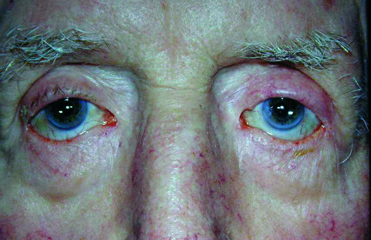
Results: All patients had a good cosmetic result. One patient had a recurrence of the upper lid lesion. Two patients complained of corneal irritation from lanugo hairs. The technique was modified to prevent this complication.
Conclusions: Large upper lid marginal defects can be readily repaired using the technique described with local advancement flaps with no significant complications.
Figures


Similar articles
-
A technique for the reconstruction of lower eyelid marginal defects.Br J Ophthalmol. 2007 Dec;91(12):1695-7. doi: 10.1136/bjo.2007.123075. Br J Ophthalmol. 2007. PMID: 18024813 Free PMC article.
-
Modified Hughes procedure for reconstruction of large full-thickness lower eyelid defects following tumor resection.Eur J Med Res. 2016 Jun 30;21(1):27. doi: 10.1186/s40001-016-0221-1. Eur J Med Res. 2016. PMID: 27364344 Free PMC article.
-
The Cutler-Beard flap for upper eyelid reconstruction: Surgical indications revisited.Eur J Ophthalmol. 2024 Nov;34(6):1795-1802. doi: 10.1177/11206721241234417. Epub 2024 Feb 21. Eur J Ophthalmol. 2024. PMID: 38384118
-
Eyelid Reconstruction: An Algorithm Based on Defect Location.J Craniofac Surg. 2022 May 1;33(3):821-826. doi: 10.1097/SCS.0000000000008433. Epub 2021 Dec 29. J Craniofac Surg. 2022. PMID: 34967774 Review.
-
Advancements in the repair of large upper eyelid defects: A 10-year review.Orbit. 2021 Dec;40(6):470-480. doi: 10.1080/01676830.2020.1820045. Epub 2020 Sep 29. Orbit. 2021. PMID: 32990145 Free PMC article. Review.
Cited by
-
Lateral eyelid rotation flap: a novel technique for reconstruction of full thickness eyelid defect.Int Ophthalmol. 2015 Dec;35(6):793-9. doi: 10.1007/s10792-015-0047-9. Epub 2015 Feb 12. Int Ophthalmol. 2015. PMID: 25673519 Review.
-
Reconstruction of the upper eyelid with flaps and free grafts after excision of Basal cell carcinoma.Case Rep Ophthalmol. 2011 Sep;2(3):347-53. doi: 10.1159/000334674. Epub 2011 Nov 15. Case Rep Ophthalmol. 2011. PMID: 22128284 Free PMC article.
-
MR-Eye: High-Resolution Microscopy Coil MRI for the Assessment of the Orbit and Periorbital Structures, Part 2: Clinical Applications.AJNR Am J Neuroradiol. 2021 Jul;42(7):1184-1189. doi: 10.3174/ajnr.A7080. Epub 2021 Mar 18. AJNR Am J Neuroradiol. 2021. PMID: 33737269 Free PMC article. Review.
-
A technique for the reconstruction of lower eyelid marginal defects.Br J Ophthalmol. 2007 Dec;91(12):1695-7. doi: 10.1136/bjo.2007.123075. Br J Ophthalmol. 2007. PMID: 18024813 Free PMC article.
-
Reconstruction of large upper eyelid defect with two composite lid margin grafts.Middle East Afr J Ophthalmol. 2010 Apr;17(2):161-4. doi: 10.4103/0974-9233.63083. Middle East Afr J Ophthalmol. 2010. PMID: 20616924 Free PMC article.
References
-
- Cook BE Jr, Bartley GB. Epidemiologic characteristics and clinical course of patients with malignant eyelid tumours in an incidence cohort in Olmstead County, Minnesota. Ophthalmology 1999;106:746–50. - PubMed
-
- Cutler NL, Beard C. A method for partial and total upper lid reconstruction. Am J Ophthalmol 1955;39:1–7. - PubMed
-
- Mustard JC. Repair and reconstruction in the orbital region. Edinburgh: Livingstone, 1966:198–201.
-
- Tenzel RR, Stewart WB. Eyelid reconstruction by semicircular flap technique. Trans Am Soc Ophthalmol Otolaryngol 1978;85:1165. - PubMed
MeSH terms
LinkOut - more resources
Full Text Sources