High resolution magnetic resonance imaging of retinoblastoma
- PMID: 12598449
- PMCID: PMC1771559
- DOI: 10.1136/bjo.87.3.330
High resolution magnetic resonance imaging of retinoblastoma
Abstract
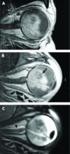
Background/aims: Diagnosis of retinoblastoma is mainly based on indirect ophthalmoscopy, but additional imaging techniques are indispensable for the staging of the disease. A new high resolution magnetic resonance imaging (MRI) technique for the examination of the eye was evaluated. A new surface coil with a diameter of 5 cm allows a field of view of 60 mm with an in-plane resolution of 0.8 mm. We compared preoperative MRI scans with the histology after enucleation in 21 cases of retinoblastoma. Parameters studied were appearance of retinoblastoma, choroidal and scleral infiltration, extraocular extension, optic nerve infiltration, and vitreous seeding.
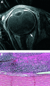
Results: All retinoblastomas could be visualised as hypointense to vitreous on T2 weighted images and slightly hyperintense to vitreous on plain T1 weighted images with a moderate enhancement after contrast application. Histology revealed seven cases with infiltration of the optic disc or optic nerve. Preoperative MRI scans depict juxtapapillary tumour masses, but it was impossible to differentiate between a juxtapapillary retinoblastoma, a prelaminar infiltration of the optic disc, or a just postlaminar optic nerve infiltration. In five of 14 cases with a proved tumour infiltration of the choroid, MRI scans showed an inhomogeneous contrast enhancement of the choroid in enhanced T1 weighted sequences beneath the retinoblastoma. Whether this sign is specific for a choroidal infiltration or is just an artefact remains unclear. High resolution MRI scans did not allow the exclusion of this form of intraocular tumour extension. All nine cases with proved vitreous seeding were not detected by MRI scans. None of these cases showed scleral infiltration or orbital tumour extension. Therefore, it is not possible to judge the rank of this technique in detecting orbital tumour growth.
Conclusion: The new MRI technique is of limited value in visualisation of prelaminar or postlaminar infiltration of the optic nerve. Advanced choroidal infiltration might be visualised by contrast enhanced T1 weighted MRI scans, but the available spatial resolution did not allow the exclusion this critical form of tumour growth by MRI scans. Nevertheless, high resolution MRI with the new surface coil has superior contrast and spatial resolution compared to computed tomograph (CT) or other available imaging techniques. MRI cannot replace CT in detecting tumour calcification but with increasing experience with this new technique it should be possible to renounce CT scans in the majority of cases of retinoblastoma.
Figures


References
-
- Eng C, Li FP, Abramson DH, et al. Mortality from second tumors among long-term survivors of retinoblastoma. J Natl Cancer Inst 1993;85:1121–8. - PubMed
-
- Shields CL, Shields JA, De Potter P, et al. Plaque radiotherapy in the management of retinoblastoma. Use as a primary and secondary treatment. Ophthalmology 1993;100:216–24. - PubMed
-
- Shields JA, Shields CL, De Potter P. Cryotherapy for retinoblastoma. Int Ophthalmol Clin 1993;33:101–5. - PubMed
-
- Shields JA, Shields CL, Parsons H, et al. The role of photocoagulation in the management of retinoblastoma. Arch Ophthalmol 1990;108:205–8. - PubMed
-
- Shields CL, Shields JA, Kiratli H, et al. Treatment of retinoblastoma with indirect ophthalmoscope laser photocoagulation. J Pediatr Ophthalmol Strabismus 1995;32:317–22. - PubMed
MeSH terms
LinkOut - more resources
Full Text Sources
Other Literature Sources
Medical
