. 2003 May 2;278(18):16347-55.
doi: 10.1074/jbc.M300259200. Epub 2003 Feb 21.

Recruitment and activation of phospholipase Cgamma1 by vascular endothelial growth factor receptor-2 are required for tubulogenesis and differentiation of endothelial cells

Affiliations

Recruitment and activation of phospholipase Cgamma1 by vascular endothelial growth factor receptor-2 are required for tubulogenesis and differentiation of endothelial cells

Rosana D Meyer et al. J Biol Chem. .

Erratum in

  • J Biol Chem. 2005 Jul 8;280(27):25948

Abstract

Vascular endothelial growth factor-mediated angiogenic signal transduction relay is achieved by coordinated induction of endothelial cell proliferation, migration, and differentiation. These complex cellular processes are most likely controlled by activation of both cooperative and antagonistic signals by vascular endothelial growth factor receptors (VEGFRs). Here, we investigated the contribution of tyrosine-phosphorylated residues of VEGFR-2/fetal liver kinase-1 to endothelial cell proliferation and differentiation and activation of signaling proteins. Mutation of tyrosine 1006 of VEGFR-2 to phenylalanine severely impaired the ability of this receptor to stimulate endothelial cell differentiation and tubulogenesis. Paradoxically, the mutant receptor stimulated endothelial cell proliferation far better than the wild-type receptor. Further analysis showed that tyrosine 1006 is responsible for phospholipase Cgamma1 (PLCgamma1) activation and intracellular calcium release in endothelial cells. Activation of PLCgamma1 was selectively mediated by tyrosine 1006. Mutation of tyrosines 799, 820, 949, 994, 1080, 1173, and 1221 had no measurable effect on the ability of VEGFR-2 to stimulate PLCgamma1 activation. Association of VEGFR-2 with PLCgamma1 was mainly established between tyrosine 1006 and the C-terminal SH2 domain of PLCgamma1 in vitro and in vivo. Taken together, the results indicate that phosphorylation of tyrosine 1006 is essential for VEGFR-2-mediated PLCgamma1 activation, calcium flux, and cell differentiation. More importantly, VEGFR-2-mediated endothelial cell proliferation is inversely correlated with the ability of VEGFR-2 to associate with and activate PLCgamma1.


Figures
F<sc>ig</sc>. 1
Fig. 1
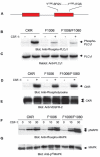
Role of individual tyrosine residues of VEGFR-2 in PLCγ1 activation. Serum-starved semiconfluent PAE cells expressing CKR were either non-stimulated or stimulated with 40 ng/ml CSF-1 for the indicated times, washed, and lysed, and cell extracts were normalized for protein. Total cell lysates were subjected to Western blot analysis using anti-phospho-PLCγ1 antibody (A). The same membrane was reprobed with anti-PLCγ1 antibody (B). A schematic representation of tyrosine residues located in the cytoplasmic region of VEGFR-2 is shown in C. Serum-starved PAE cells expressing wild-type CKR, F799/CKR, or F1173/CKR were treated with CSF-1 for 10 min, washed, lysed, and subjected to Western blot analysis using anti-phospho-PLCγ1 (D). The same membrane was reprobed with anti-PLCγ1 antibody (E). Serum-starved PAE cells expressing wild-type CKR, F820/CKR, F949/CKR, F994/CKR, F1006/CKR, or F1221/CKR were treated with CSF-1, and total cell lysates were resolved by SDS-PAGE and blotted with anti-phospho-PLCγ1 antibody (F). The same membrane was reprobed with anti-PLCγ1 antibody for protein levels (G).
F<sc>ig</sc>. 2
Fig. 2
Mutation of tyrosine 1006 impairs the ability of VEGFR-2 to stimulate PLCγ1 activation, but not MAPK activation. Shown is a schematic representation of tyrosines 1006 and 1080 of VEGFR-2 (A). Serum-starved semiconfluent PAE cells expressing CKR, F1006/CKR, F1006/F1080/CKR were either non-stimulated or stimulated with 40 ng/ml CSF-1 for 10 min, washed, and lysed, and cell extracts were normalized for protein. Total cell lysates were subjected to Western blot analysis using anti-phospho-PLCγ1 antibody (B). The same membrane was reprobed with anti-PLCγ1 antibody (C). Serum-starved cells were stimulated with CSF-1 as described for B, immuno-precipitated with anti-VEGFR-2 antibody, and subjected to Western blot analysis using anti-phosphotyrosine antibody (D). The same membrane was reprobed with anti-VEGFR-2 antibody (E). Serum-starved cells were stimulated with CSF-1 as described for B, and total cell lysates were subjected to Western blot analysis using anti-phospho-MAPK (p44/42) antibody (F). The same membrane was reprobed with anti-MAPK antibody (G).
F<sc>ig</sc>. 3
Fig. 3
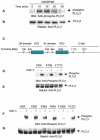
The C-SH2 domain of PLCγ1 associates with tyrosine 1006 of VEGFR-2. Shown is a schematic presentation of PLCγ1 and GST fusion proteins containing the SH2 domains of PLCγ (A). Serum-starved semiconfluent PAE cells expressing CKR were stimulated with 40 ng/ml CSF-1, washed, and lysed, and cell extracts were normalized for protein. Total cell lysates were incubated with Sepharose-bound GST-N-SH2, GST-C-SH2, or GST-N+C-SH2. After extensive washing, the precipitated proteins were resolved by SDS-PAGE and immunoblotted with anti-VEGFR-2 antibody (B). Serum-starved semiconfluent PAE cells expressing CKR, F1006/CKR, and F1173/CKR were stimulated with 40 ng/ml CSF-1, washed, and lysed, and cell extracts were normalized for protein. Total cell lysates were incubated with either GST-C-SH2 or GST-C+N-SH2 as described for B and subjected to Western blot analysis using anti-VEGFR-2 antibody (C).
F<sc>ig</sc>. 4
Fig. 4
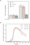
The presence of tyrosine 1006 of VEGFR-2 is required for inositol phosphate production and calcium flux. PAE cells expressing CKR, F1173/CKR, and F1006/CKR were incubated with 1μCi/ml myo-[H]inositol for 48 h in DMEM supplemented with 0.1% bovine serum albumin, and phospholipid production was measured by scintillation counting as described under “Materials and Methods” (A). Serum-starved PAE cells expressing CKR, F1173/CKR, and F1006/CKR were grown on glass coverslips and stimulated with CSF-1, and calcium flux was measured by confocal microscopy using Fluo-3/AM as a probe as described under “Materials and Methods” (B). IPs, inositol phosphates.
F<sc>ig</sc>. 5
Fig. 5
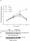
Mutation of tyrosine 1006 enhances the ability of VEGFR-2 to stimulate cell proliferation. Serum-starved PAE cells expressing wild-type CKR and tyrosine mutant CKRs were treated with different concentrations of CSF-1, and DNA synthesis was measured by [3H]thymidine uptake. The results are expressed as the mean (cpm/well) ± S.D. of quadruplicates. The data are expressed as a ratio of stimulated versus non-stimulated samples (A). Serum-starved semiconfluent PAE cells expressing CKR or F1006/CKR were either non-stimulated or stimulated with 40 ng/ml CSF-1 for 20 or 30 min, washed, and lysed, and cell extracts were normalized for protein. Total cell lysates were subjected to Western blot analysis using anti-phospho-AKT antibody (B). The same membrane was reprobed with anti-AKT antibody (C).
F<sc>ig</sc>. 6
Fig. 6
Mutation of tyrosine 1006 abrogates the ability of VEGFR-2 to stimulate differentiation and tubulogenesis of PAE cells. PAE cells expressing CKR, F1173/CKR, and F1006/CKR were plated in six-well plates, serum-starved overnight, and either non-stimulated or stimulated with CSF-1 (20 ng/ml) for 24–30 h. Morphological changes associated with CSF-1 stimulation were viewed under an inverted microscope (magnification ×10) and photographed with the SPOT camera system (A). PAE cells expressing CKR, F1173/CKR, and F1006/CKR were prepared as spheroids and subjected to an in vitro angiogenesis/tubulogenesis assay as described under “Materials and Methods.” Sprouting and tubulogenesis were observed after 2 days under an inverted phase-contrast microscope, and pictures were taken using the SPOT camera system (B).
F<sc>ig</sc>. 7
Fig. 7
Inhibition of PLCγ1 by pharmacological means inhibits the ability of CKR to stimulate tubulogenesis. PAE cells expressing CKR were prepared in spheroid forms and subjected to an in vitro angiogenesis/tubulogenesis assay as described for Fig. 6B. Spheroids were treated with different concentrations of the PLCγ1 inhibitor U73122 as indicated. Sprouting and tubulogenesis were observed after 2 days under an inverted phase-contrast microscope, and pictures were taken using the SPOT camera system.

References

    1. Risau W. Nature. 1997;386:671–674. - PubMed
    1. Folkman J, D'Amore PA. Cell. 1996;87:1153–1155. - PubMed
    1. Takahashi T, Yamaguchi S, Chida K, Shibuya M. EMBO J. 2001;20:2768–2778. - PMC - PubMed
    1. Dayanir V, Meyer RD, Lashkari K, Rahimi N. J. Biol. Chem. 2001;276:17686–17692. - PubMed
    1. Cunningham SA, Arrate MP, Brock TA, Waxham MN. Biochem. Biophys. Res. Commun. 1997;240:635–639. - PubMed

Publication types

MeSH terms

Substances