. 2003 Apr 1;548(Pt 1):111-20.
doi: 10.1113/jphysiol.2002.033100. Epub 2003 Feb 21.

Alpha 1-adrenoceptor-activated cation currents in neurones acutely isolated from rat cardiac parasympathetic ganglia

Affiliations

Alpha 1-adrenoceptor-activated cation currents in neurones acutely isolated from rat cardiac parasympathetic ganglia

Hitoshi Ishibashi et al. J Physiol. .

Abstract

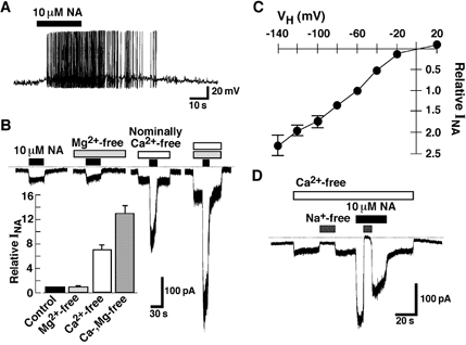
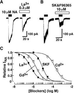
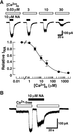
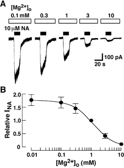
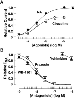
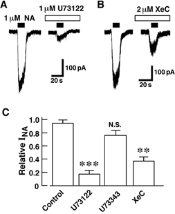
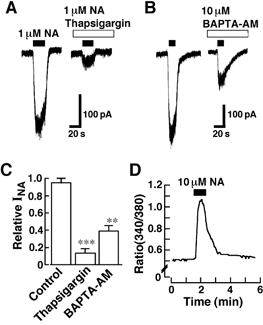
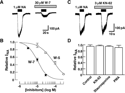
The noradrenaline (NA)-induced cation current was investigated in neurones freshly isolated from rat cardiac parasympathetic ganglia using the nystatin-perforated patch recording configuration. Under current-clamp conditions, NA depolarized the membrane, eliciting repetitive action potentials. NA evoked an inward cation current under voltage-clamp conditions at a holding potential of -60 mV. The NA-induced current was inhibited by extracellular Ca2+ or Mg2+, with a half-maximal concentration of 13 microM for Ca2+ and 1.2 mM for Mg2+. Cirazoline mimicked the NA response, and prazosin and WB-4101 inhibited the NA-induced current, suggesting the contribution of an alpha1-adrenoceptor. The NA-induced current was inhibited by U73122, a phospholipase C (PLC) inhibitor. The membrane-permeable IP3 receptor blocker xestospongin-C also blocked the NA-induced current. Furthermore, pretreatment with thapsigargin and BAPTA-AM could inhibit the NA response while KN-62, phorbol 12-myristate 13-acetate (PMA) and staurosporine had no effect. These results suggest that NA activates the extracellular Ca2+- and Mg2+-sensitive cation channels via alpha 1-adrenoceptors in neurones freshly isolated from rat cardiac parasympathetic ganglia. This activation mechanism also involves phosphoinositide breakdown, release of Ca2+ from intracellular Ca2+ stores and calmodulin. The cation channels activated by NA may play an important role in neuronal membrane depolarization in rat cardiac ganglia.


Figures

Figure 1
Figure 1. Noradrenaline response in isolated cardiac ganglion neurones
A, depolarization of a rat cardiac ganglion neurone by noradrenaline (NA). Recordings were made under current-clamp conditions. Horizontal filled bar above the trace indicates the application of NA. The figure is representative of 6 reproducible experiments. B, inward currents evoked by NA (INA). All current traces were recorded from the same neurone. Holding potential (VH) was −60 mV. The NA-induced current was sensitive to extracellular divalent cations. Dotted line shows the zero current level. The inset summarizes the effects of Ca2+ and Mg2+ on the NA-induced current. The current amplitude was normalized to that observed in the normal external solution containing 2 mm Ca2+ and 1 mm Mg2+. Each column shows the mean of 5–7 experiments. C, current-voltage relationship of the NA (10 μm)-induced current. The amplitude of the NA-induced current at various VH was normalized to that observed at −60 mV in each neurone. Each point is the mean of 4–6 experiments. Vertical bars indicate ±s.e.m.D, extracellular Na+ dependency. The current trace is representative of 4 reproducible experiments.
Figure 2
Figure 2. Effects of organic and inorganic cation channel blockers
Experiments were performed in Ca2+-free solution containing 1 mm Mg2+. Each blocker was applied 1 min before the application of NA. A, effect of La3+ on the NA-induced current. B, effect of SK&F96365. C, concentration-dependent inhibition of the NA-induced current by La3+, Gd3+, SK&F96365 and Cd2+. Curves are the best non-linear fits to the Hill equation, 1/1 + (C/Ki) nH where C, Ki and nH denote the concentration of each blocker applied, dissociation constant and Hill coefficient, respectively. Each point is the mean of 6–8 experiments. Vertical bars indicate ±s.e.m.
Figure 3
Figure 3. Effects of [Ca2+]o on the NA-induced current
Experiments were performed in the solution containing 1 mm Mg2+. A, the effect of [Ca2+]o on NA-induced current. All current traces were obtained from the same neurone. NA was applied every 6 min in various [Ca2+]o. Lower panel shows the [Ca2+]o dependency. Current amplitude was normalized to that observed in the solution containing 30 nm Ca2+. Each point is the mean of 4–6 experiments. Vertical bars indicate ±s.e.m. B, rapid inhibition of the NA-induced current by extracellular Ca2+. During the application of NA in Ca2+-free solution, the switch of extracellular solution to that containing 2 mm Ca2+ rapidly inhibited the current within 1 s The figure is representative of 5 reproducible experiments.
Figure 4
Figure 4. Effects of extracellular Mg2+ concentration
A, representative current traces induced by NA in the solution containing various concentrations of Mg2+. In this experiment, Mg2+ was simply added to the nominally divalent cation-free solution. NA was applied every 6 min. B, concentration-inhibition curve for the NA-induced current by Mg2+ in divalent cation-free extracellular solution. The amplitude of the NA-induced current in various concentrations of Mg2+ was normalized to that in the solution containing 1 mm Mg2+. Each point is the mean of 4–5 experiments. Vertical bars indicate ±s.e.m.
Figure 5
Figure 5. Effects of adrenoceptor agonists and antagonists
A, concentration-response curve for NA and cirazoline. The current evoked by various concentrations of NA and cirazoline was normalized to that induced by 10 μm NA. In this and subsequent figures, electrophysiological experiments were performed at a VH of −60 mV in the Ca2+-free solution containing 2 mm EGTA and 1 mm Mg2+. Each point is the mean of 4–5 experiments. Vertical bars indicate ±s.e.m.B, effects of prazosin, WB-4101 and yohimbine on the current induced by 10 μm NA. These antagonists were applied 1 min before the application of NA. Each point is the mean of 4–6 experiments. Vertical bars indicate ±s.e.m.
Figure 6
Figure 6. Contribution of PLC and IP3 receptors
A and B, inhibitory effects of PLC inhibitor U73122 (A) and membrane-permeable IP3 receptor antagonist XeC (B) on the NA-induced current. Each inhibitor was applied 4 min before the application of NA. C, effects of 1 μm U73122, 1 μm U73343 and 2 μm XeC. Current responses in the presence of various inhibitors were normalized to that obtained just before the application of each inhibitor. Each column represents the mean of 4–6 experiments. **P < 0.01 vs. control. ***P < 0.001 vs. control. N.S., no significant difference from control.
Figure 7
Figure 7. Involvement of Ca2+ release from intracellular Ca2+ stores in the NA response
A and B, representative current traces showing the inhibitory effects of thapsigargin and BAPTA-AM on the NA response. Thapsigargin and BAPTA-AM were applied 4 min before the application of NA. C, inhibitory effects of thapsigargin and BAPTA-AM on the NA-induced current. Each column represents the mean of 5–6 experiments. **P < 0.01 vs. Control. ***P < 0.001 vs. Control. D, increase in [Ca2+]i (measured as 340 nm/380 nm fura-2 fluorescence ratio) induced by NA even in the absence of extracellular Ca2+. The response is representative of 8 reproducible experiments.
Figure 8
Figure 8. Effects of calmodulin inhibitor, KN-62 and PKC modulators on the NA-induced current
A, inhibitory effect of W-7 on the NA-induced current. B, concentration-inhibition curve of W-7 and W-5 for the NA (1 μm)-induced current. W-7 and W-5 were applied 4 min before the application of NA. Each point is the mean of 5 experiments. Vertical bars indicate ±s.e.m. C, KN-62 had no effect on the NA response. D, effects of 3 μm KN-62, 0.3 μm staurosporine and 0.3 μm PMA on the NA-induced current. KN-62, staurosporine and PMA were applied 10 min before the application of NA.

Similar articles

Cited by

References

    1. Akasu T, Gallagher JP, Nakamura T, Shinnick-Gallagher P, Yoshimura M. Noradrenaline hyperpolarization and depolarization in cat vesical parasympathetic neurons. J Physiol. 1985;361:165–184. - PMC - PubMed
    1. Albert AP, Large WA. A Ca2+-permeable non-selective cation channel activated by depletion of internal Ca2+ stores in single rabbit portal vein myocytes. J Physiol. 2002;538:717–728. - PMC - PubMed
    1. Ardell JL, Randall WC. Selective vagal innervation of sinoatrial and atrioventricular nodes in canine heart. Am J Physiol. 1986;251:H764–773. - PubMed
    1. Arima J, Kubo C, Ishibashi H, Akaike N. α2-Adrenoceptor-mediated potassium currents in acutely dissociated rat locus coeruleus neurones. J Physiol. 1998;508:57–66. - PMC - PubMed
    1. Beaudet MM, Parsons RL, Braas KM, May V. Mechanisms mediating pituitary adenylate cyclase-activating polypeptide depolarization of rat sympathetic neurons. J Neurosci. 2000;20:7353–7361. - PMC - PubMed

Publication types

MeSH terms

LinkOut - more resources