

Damage to developing mouse skeletal muscle myotubes in culture: protective effect of heat shock proteins
- PMID: 12598587
- PMCID: PMC2342900
- DOI: 10.1113/jphysiol.2002.034520
Damage to developing mouse skeletal muscle myotubes in culture: protective effect of heat shock proteins
Abstract
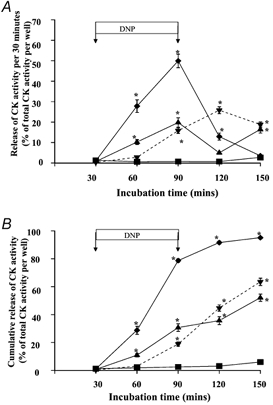
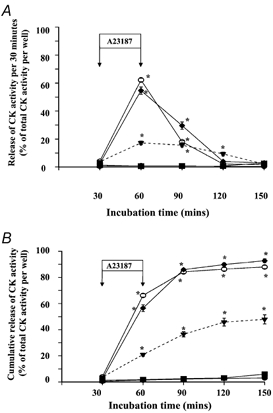
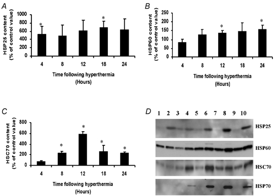
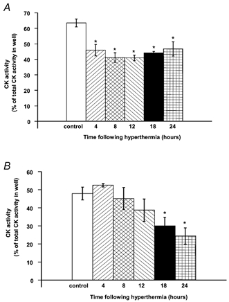
Damage to skeletal muscle occurs following excessive exercise, upon reperfusion following ischaemia and in disease states, such as muscular dystrophy. Key mechanisms by which damage is thought to occur include a loss of intracellular calcium homeostasis, loss of energy supply to the cell, increased activity of oxidising free radical-mediated reactions and activation of apoptosis pathways. An increased cellular content of heat shock proteins (HSPs) has been shown to protect skeletal muscle against some forms of damage, although the mechanistic basis of this protection is not clearly understood. The aim of this study was to establish a cell culture-based model of damage to C2C12 skeletal muscle cells using the calcium ionophore, A23187 and the mitochondrial uncoupler, 2,4-dinitrophenol (DNP) as damaging agents. Treatment of cells with 1 mM DNP for 60 min resulted in the release of 63.5 % of intracellular creatine kinase (CK) activity over the 3 h experimental period. Treatment of cells with 10 microM A23187 for 30 min resulted in the release of 47.9 % of CK activity. Exposure of myotubes to a period of hyperthermia resulted in a significant increase in their content of HSP25, HSP60, HSC70 (heat shock cognate) and HSP70. This increase in HSPs was associated with significant protection against both DNP-induced and A23187-induced damage to the myotubes. These results indicate that an increased content of HSPs may provide protection against the muscle damage that occurs by a pathological increase in intracellular calcium or uncoupling of the mitochondrial respiratory chain.
Figures

Similar articles
-
The exercise-induced stress response of skeletal muscle, with specific emphasis on humans.Sports Med. 2009;39(8):643-62. doi: 10.2165/00007256-200939080-00003. Sports Med. 2009. PMID: 19769414 Review.
-
Glutathione depletion during experimental damage to rat skeletal muscle and its relevance to Duchenne muscular dystrophy.Clin Sci (Lond). 1991 Jun;80(6):559-64. doi: 10.1042/cs0800559. Clin Sci (Lond). 1991. PMID: 1647917
-
AICAR-induced activation of AMPK negatively regulates myotube hypertrophy through the HSP72-mediated pathway in C2C12 skeletal muscle cells.Am J Physiol Endocrinol Metab. 2014 Feb;306(3):E344-54. doi: 10.1152/ajpendo.00495.2013. Epub 2013 Dec 17. Am J Physiol Endocrinol Metab. 2014. PMID: 24347059
-
Acute heat stress prior to downhill running may enhance skeletal muscle remodeling.Cell Stress Chaperones. 2012 Nov;17(6):693-705. doi: 10.1007/s12192-012-0343-5. Epub 2012 May 17. Cell Stress Chaperones. 2012. PMID: 22589083 Free PMC article.
-
Changes in skeletal muscle heat shock proteins: pathological significance.Front Biosci. 2001 Jan 1;6:D12-25. doi: 10.2741/liu. Front Biosci. 2001. PMID: 11145923 Review.
Cited by
-
MicroRNA-216b inhibits heat stress-induced cell apoptosis by targeting Fas in bovine mammary epithelial cells.Cell Stress Chaperones. 2018 Sep;23(5):921-931. doi: 10.1007/s12192-018-0899-9. Epub 2018 May 5. Cell Stress Chaperones. 2018. PMID: 29730848 Free PMC article.
-
Diathermy treatment increases heat shock protein expression in female, but not male skeletal muscle.Eur J Appl Physiol. 2008 Feb;102(3):319-23. doi: 10.1007/s00421-007-0572-y. Epub 2007 Oct 18. Eur J Appl Physiol. 2008. PMID: 17943309
-
Living at high altitude in combination with sea-level sprint training increases hematological parameters but does not improve performance in rats.Eur J Appl Physiol. 2011 Jun;111(6):1147-56. doi: 10.1007/s00421-010-1740-z. Epub 2010 Dec 1. Eur J Appl Physiol. 2011. PMID: 21120517
-
Elevation in heat shock protein 72 mRNA following contractions in isolated single skeletal muscle fibers.Am J Physiol Regul Integr Comp Physiol. 2008 Aug;295(2):R642-8. doi: 10.1152/ajpregu.00852.2007. Epub 2008 Jun 4. Am J Physiol Regul Integr Comp Physiol. 2008. PMID: 18525012 Free PMC article.
-
Evidence for ACTN3 as a genetic modifier of Duchenne muscular dystrophy.Nat Commun. 2017 Jan 31;8:14143. doi: 10.1038/ncomms14143. Nat Commun. 2017. PMID: 28139640 Free PMC article.
References
-
- Diedrichs F, Muhlhaus K, Trautschold I. On the mechanism of lactate dehydrogenase release from skeletal muscle in relation to the control of cell volume. Enzyme. 1979;24:404–415. - PubMed
-
- Faulkner JA, Brooks SV. Muscle damage induced by contraction: in an in situ single skeletal muscle model. In: Salmons S, editor. Muscle Damage. Oxford: Oxford University Press; 1997. pp. 28–40.
-
- Faulkner JA, Brooks SV, Opiteck JA. Injury to skeletal muscle fibres during contractions: conditions of occurrence and prevention. Phys Ther. 1993;73:911–921. - PubMed
Publication types
MeSH terms
Substances
Grants and funding
LinkOut - more resources
Full Text Sources
Other Literature Sources
Research Materials
Miscellaneous