. 2003 Feb 15;23(4):1119-24.
doi: 10.1523/JNEUROSCI.23-04-01119.2003.

The role of synaptic GTPase-activating protein in neuronal development and synaptic plasticity

Affiliations

The role of synaptic GTPase-activating protein in neuronal development and synaptic plasticity

Jee Hae Kim et al. J Neurosci. .

Abstract

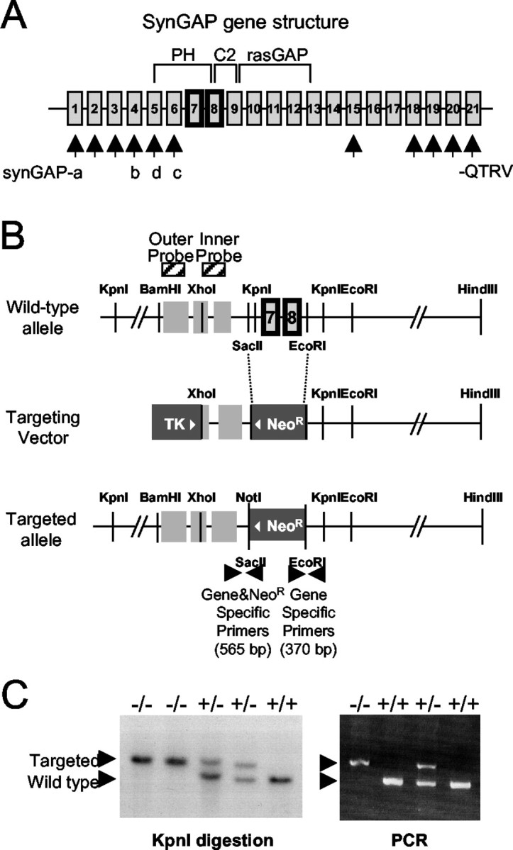
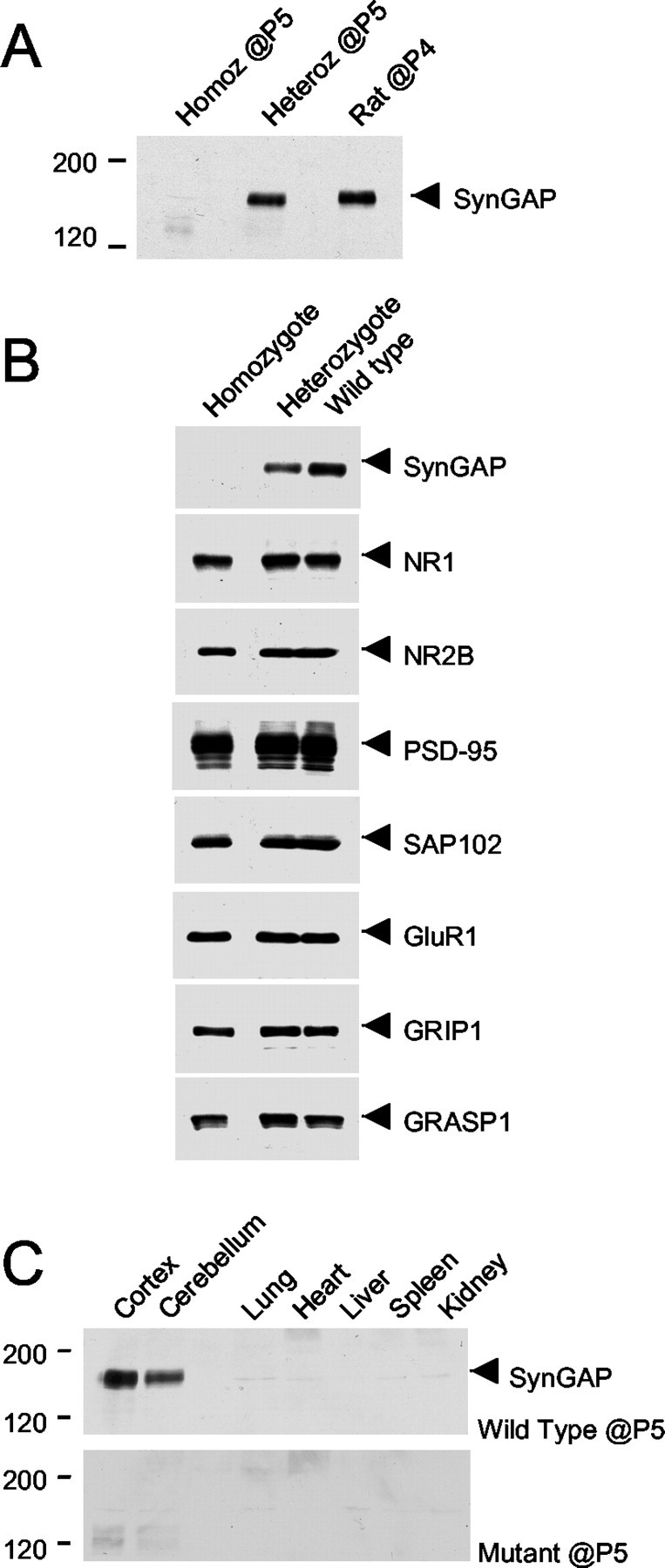
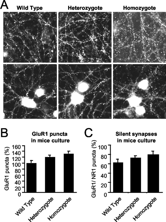
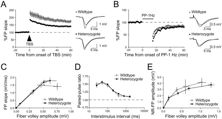
Synaptic GTPase-activating protein (SynGAP) is a neuronal RasGAP (Ras GTPase-activating protein) that is selectively expressed in brain and highly enriched at excitatory synapses, where it negatively regulates Ras activity and its downstream signaling pathways. To investigate the physiological role of SynGAP in the brain, we have generated mutant mice lacking the SynGAP protein. These mice exhibit postnatal lethality, indicating that SynGAP plays a critical role during neuronal development. In addition, cell biological experiments show that neuronal cultures from mutant mice have more synaptic AMPA receptor clusters, suggesting that SynGAP regulates glutamate receptor synaptic targeting. Moreover, electrophysiological studies demonstrated that heterozygous mutant mice have a specific defect in hippocampal long-term potentiation (LTP). These studies show that the regulation of synaptic Ras signaling by SynGAP is important for proper neuronal development and glutamate receptor trafficking and is critical for the induction of LTP.


Figures

Fig. 1.
Fig. 1.
SynGAP splice variants and domain structure and gene targeting strategy. A, N-terminal splicing leads to different start sites and sequences in SynGAP-a, -b, -c, and -d. SynGAP protein contains a pleckstrin homology (PH) domain, a phospholipid-dependent Ca2+ binding motif (C2) domain, a Ras GTPase-activation protein (RasGAP) domain, and a C-terminal sequence PSD-95/discs large/zona occludens-1 domain binding motif (QTRV). Alternative splicing occurs also at the C terminal with C-terminal sequences other than −QTRV.B, The SynGAP gene structure is shown (not drawn to scale) of the region analyzed for gene targeting. Targeting of the SynGAP gene was performed by replacing the SacII andEcoRI fragments of the SynGAP gene containing two exons with the neoR cassette. The targeting construct spanning the XhoI and HindIII fragment of the SynGAP gene is 11.5 kb long. Outer and inner probes were used for Southern blotting. The PCR primers used for genotyping and the predicted amplified sizes are shown. TK, Thymidine kinase. C, Genotype analyses of tail DNA of second filial generation (F2) mice by PCR and Southern blotting. The wild-type allele is detected by Southern blotting after digestion with the KpnI restriction enzyme (left). The size of the detected wild-type band is 1.8 kb longer than that of the targeted allele. Two alleles can be distinguished using PCR primer sets (right).
Fig. 2.
Fig. 2.
A proper expression of SynGAP protein is abolished in the mutant mice, whereas other synaptic proteins are not affected at P5. A, Mouse brain homogenates were prepared and immunoblotted with the anti-GAP SynGAP antibodies at P5 and compared with that of rat brain homogenate at P4. With an equal amount of protein loaded in each lane, SynGAP protein expression is absent in the sample from a homozygous (Homoz) mouse.Heteroz, Heterozygous. B, The expression of proteins at synapses was examined in the SynGAP mice using the antibodies to the proteins indicated, and no detectable change in the expression was seen. C, Tissue distribution of SynGAP protein in the mutant and the wild-type mice at P5 was examined using the α-GAP domain antibody. In wild-type mice, a prominent band of ∼130 kDa was detected in the cortex and the cerebellum. In contrast, the ∼130 kDa protein was not detected in the homozygotes or in non-neuronal tissues.
Fig. 3.
Fig. 3.
The number of AMPA receptor clusters in the SynGAP mutant mice is increased. A, Primary cortical cultures from the SynGAP mutant mice and their wild-type and heterozygous littermates were immunostained with anti-GluR1 antibodies after 18–20 DIV. There was an increase in the number of GluR1-positive clusters in the cultures prepared from the homozygous pups. B, Quantitation of GluR1-positive puncta in SynGAP mouse neuronal cultures at 18–20 DIV (n = 14, n = 19, and n = 13, respectively; p < 0.05; ANOVA; F = 3.52). C, The number of morphological silent synapses in the cultures was quantitated by comparing the number of AMPA receptor cluster/NMDA receptor cluster puncta (n = 9, n = 13, andn = 7, respectively) at 18–20 DIV.
Fig. 4.
Fig. 4.
A, Schaffer collateral to CA1 LTP in adult SynGAP heterozygotes (●; n = 20 slices from 5 animals) are significantly reduced compared with wild-type littermates (○; n = 16 slices from 5 animals). FP traces taken just before and 1 hr after TBS for wild types and heterozygotes are shown to the right. B, No significant difference in PP-LTD (PP-1Hz) in SynGAP heterozygotes (●; n = 21 slices from 4 animals) and wild types (○; n = 18 slices from 4 animals). FP traces taken just before and 1 hr after the initiation of PP-1 Hz are shown to the right. C, AMPA receptor-mediated synaptic transmission measured as the initial FP slope plotted against fiber volley amplitude. Plots of both wild types (○; n = 32 slices from 8 animals) and heterozygotes (●; n = 33 slices from 8 animals) essentially overlap, suggesting that synaptic transmission is normal in heterozygotes. D, No difference was observed in presynaptic function as monitored by paired-pulse facilitation between wild types (○; n = 14 slices from 5 animals) and heterozygotes (●; n = 14 slices from 5 animals). Paired pulses were given at interstimulus intervals of 25, 50 100, 200, 400, 800, and 1600 msec at baseline stimulus intensity.E, Pharmacologically isolated NMDA receptor-mediated synaptic transmission does not differ much between SynGAP heterozygous and wild-type littermates. NMDA receptor-mediated synaptic responses were pharmacologically isolated by bath application of ACSF with 0 mm Mg2+ and 10 μm NBQX. An input–output curve was generated by plotting the amplitude of NMDA receptor (NR)-mediated FP against fiber volley amplitude. At the end of each experiment, 100 μmd,l-APV was added to the bath, completely abolishing the responses (data not shown). Dashed lines indicate normalized FP and paired-pulse facilitation ratio.

References

    1. Bliss TV, Collingridge GL. A synaptic model of memory: long-term potentiation in the hippocampus. Nature. 1993;361:31–39. - PubMed
    1. Bokoch GM, Der CJ. Emerging concepts in the Ras superfamily of GTP-binding proteins. FASEB J. 1993;7:750–759. - PubMed
    1. Brambilla R, Gnesutta N, Minichiello L, White G, Roylance AJ, Herron CE, Ramsey M, Wolfer DP, Cestari V, Rossi-Arnaud C, Grant SG, Chapman PF, Lipp HP, Sturani E, Klein R. A role for the Ras signalling pathway in synaptic transmission and long-term memory. Nature. 1997;390:281–286. - PubMed
    1. Chen HJ, Rojas-Soto M, Oguni A, Kennedy MB. A synaptic Ras-GTPase activating protein (p135 SynGAP) inhibited by CaM kinase II. Neuron. 1998;20:895–904. - PubMed
    1. Dong H, O'Brien RJ, Fung ET, Lanahan AA, Worley PF, Huganir RL. GRIP: a synaptic PDZ domain-containing protein that interacts with AMPA receptors. Nature. 1997;386:279–284. - PubMed