Comparative Study
. 2003 Feb 15;23(4):1133-41.
doi: 10.1523/JNEUROSCI.23-04-01133.2003.

Modulation of the kv3.1b potassium channel isoform adjusts the fidelity of the firing pattern of auditory neurons

Affiliations
Comparative Study

Modulation of the kv3.1b potassium channel isoform adjusts the fidelity of the firing pattern of auditory neurons

Carolyn M Macica et al. J Neurosci. .

Abstract

Neurons of the medial nucleus of the trapezoid body, which transmit auditory information that is used to compute the location of sounds in space, are capable of firing at high frequencies with great temporal precision. We found that elimination of the Kv3.1 gene in mice results in the loss of a high-threshold component of potassium current and failure of the neurons to follow high-frequency stimulation. A partial decrease in Kv3.1 current can be produced in wild-type neurons of the medial nucleus of the trapezoid body by activation of protein kinase C. Paradoxically, activation of protein kinase C increases temporal fidelity and the number of action potentials that are evoked by intermediate frequencies of stimulation. Computer simulations confirm that a partial decrease in Kv3.1 current is sufficient to increase the accuracy of response at intermediate frequencies while impairing responses at high frequencies. We further establish that, of the two isoforms of the Kv3.1 potassium channel that are expressed in these neurons, Kv3.1a and Kv3.1b, the decrease in Kv3.1 current is mediated by selective phosphorylation of the Kv3.1b isoform. Using site-directed mutagenesis, we identify a specific C-terminal phosphorylation site responsible for the observed difference in response of the two isoforms to protein kinase C activation. Our results suggest that modulation of Kv3.1 by phosphorylation allows auditory neurons to tune their responses to different patterns of sensory stimulation.


Figures
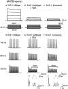
Fig. 1.
Fig. 1.
Representative traces comparing native high-threshold current from a 14 d wild-type MNTB neuron with the high-threshold current from a 14 d Kv3.1 mutant MNTB neuron.a, b, Outward currents evoked by stepping from a holding potential of −40 mV for 2 min (to ensure complete inactivation of low-threshold current) to test potentials from −80 to +40 mV in 20 mV increments in wild-type mice before and after treatment with 1 mm TEA or in Kv3.1 mutant mice (c). d, e, Representative recording from an MNTB neuron in response to brief current injections (2 nA, 0.3 msec) at three different test frequencies (100–300 Hz) in wild-type mice before and after treatment with 1 mm TEA or in Kv3.1 mutant mice (f). Bars denote failure to evoke an action potential in response to a stimulus. Failure was defined as a membrane depolarization to less than −10 mV in response to a current injection. Expanded traces below compare a successful action potential with failures, which showed little regenerative response.
Fig. 2.
Fig. 2.
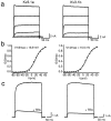
Comparison of Kv3.1 current recorded from CHO cells stably transfected with Kv3.1a or Kv3.1b. a, Whole-cell current was evoked by depolarizing the membrane from a holding potential of −80 to +60 mV in 20 mV increments.b, Normalized conductance–voltage relationship for Kv3.1a versus Kv3.1b. Conductance values (G) were obtained as described in Materials and Methods. c, Sensitivity of Kv3.1a or Kv3.1b to 1 mm TEA. Currents were evoked as described ina.
Fig. 3.
Fig. 3.
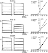
Effect of 100 nm PMA on Kv3.1 current.a, Whole-cell currents evoked from CHO cells stably expressing Kv3.1a from a holding potential of −70 mV to test potentials of −80 to +60 mV in 20 mV increments (left) and corresponding current–voltage relationship of evoked currents (right) before and after PMA treatment.b, Whole-cell currents evoked from CHO cells stably expressing Kv3.1b (left) and current–voltage relationship of evoked currents. c, Whole-cell currents evoked from CHO cells stably expressing Kv3.1b mutant S503A (left) and current–voltage relationship of evoked currents.
Fig. 4.
Fig. 4.
Time course of the effect of 100 nmPMA on Kv3.1 current amplitude. Outward current evoked from a holding potential of −70 mV in Kv3.1-transfected cells or from a holding potential of −40 mV in MNTB neurons to a test potential of +40 mV was monitored in perforated patches every 1 min in response to treatment with exogenous PMA in Kv3.1a-transfected cells (filled circles), in Kv3.1b-transfected cells (filled squares), or in MNTB neurons (filled triangles). The effect of the inactive phorbol ester 4α-PMA was also tested on Kv3.1b-transfected cells (open squares) and MNTB neurons (open triangles).
Fig. 5.
Fig. 5.
In vivo phosphorylation and phosphoamino acid analysis of Kv3.1 in CHO cells. a, CHO cells expressing Kv3.1a or Kv3.1b were radiolabeled with [32P]orthophosphate to equilibrium, stimulated with or without 100 nm PMA for 15 min, and lysed. Lysates were immunoprecipitated with Kv3.1 antibody (Kv3.1a, lanes 1 and 2, with and without PMA, respectively or Kv3.1b, lanes 4 and 5, with and without PMA, respectively) and resolved as outlined in Materials and Methods. An additional 32P-labeled Kv3.1a or Kv3.1b immunoprecipitate was treated with calf intestinal alkaline phosphatase for 1 hr at 37°C (lanes 3 and 6, respectively). b, Phosphoamino acid analysis of the Kv3.1a or Kv3.1b channel protein obtained from lysates and immunoprecipitated and electrophoresed as above. The gel was transferred to a PVDF membrane, and the Kv3.1 band was visualized by autoradiography. The excised protein was subjected to phosphoamino acid analysis as outlined in Materials and Methods and visualized by ninhydrin staining (top), and standard phosphoamino acids were visualized by autoradiography (bottom).
Fig. 6.
Fig. 6.
Molecular identity and effect of 100 nm PMA on native Kv3.1 current. a, Coimmunoprecipitation of Kv3.1a and Kv3.1b in brain homogenates or in stably transfected CHO cells. Lane 1, Kv3.1a/CHO with N-terminal antibody; lane 2, Kv3.1b/CHO with N-terminal antibody; lane 3, Kv3.1b/CHO with C-terminal antibody; lane 4, rat brain membranes with N-terminal antibody; lane 5, rat brain membranes with C-terminal antibody. b, Whole-cell currents evoked from a 13 d MNTB neuron from a holding potential of −40 mV to test potentials of −80 to +60 mV (left) and current–voltage relationship of evoked currents before and after PMA treatment.
Fig. 7.
Fig. 7.
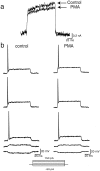
Effect of 100 nm PMA on low-threshold current from an MNTB neuron. a, Low-threshold current was obtained by subtracting the high-threshold component of outward current evoked from a holding potential of −40 mV to a test potential of −20 mV from total outward current evoked from a holding potential of −70 mV to a test potential of −20 mV (ILT =ITOTIHT) before and after PMA treatment (n = 8). b, Current-clamp recording from an MNTB neuron in response to a series of 100 msec current injections ranging from −50 to 150 pA before and after PMA treatment (n = 4).
Fig. 8.
Fig. 8.
Effect of PMA treatment on firing properties of an MNTB neuron (13 d old) in response to different frequencies of stimulation (n = 6). a, Plots of the delay from the onset of the stimulus pulse to the peak of the action potential before (Control) and 15 min after (PMA) treatment with 100 nm PMA.Arrows denote failure to evoke an action potential in response to a stimulus. Failure was defined as a membrane depolarization to less than −10 mV in response to a current injection (one that has no detectable regenerative component). b, Superimposed action potentials in response to 100, 300, and 400 Hz stimulation. Failures were omitted from the superimposedtraces.
Fig. 9.
Fig. 9.
Model of an MNTB neuron in response to different frequencies of stimulation. a, Plots of the delay from the onset of the stimulus pulse to the peak of the action potential under control conditions (150 nS Kv3.1 conductance) and under conditions in which the level of Kv3.1 current amplitude is reduced to a similar degree as in PMA-treated neurons (100 nS Kv3.1 conductance).Arrows denote failure to evoke an action potential in response to a stimulus. Failure was defined as a membrane depolarization to less than −10 mV in response to a current injection.b, Superimposed action potentials in response to 100, 350, 360, or 410 Hz stimulation. Failures were omitted from the superimposed traces.

Similar articles

Cited by

References

    1. Banks MI, Smith PH. Intracellular recordings from neurobiotin-labeled cells in brain slices of the rat medial nucleus of the trapezoid body. J Neurosci. 1992;12:2819–2837. - PMC - PubMed
    1. Borst JG, Helmchen F, Sakmann B. Pre- and postsynaptic whole-cell recordings in medial nucleus of trapezoid body of the rat. J Physiol (Lond) 1995;489:825–840. - PMC - PubMed
    1. Brew HM, Forsythe ID. Two voltage-dependent K+ conductances with complementary functions in postsynaptic integration at a central auditory synapse. J Neurosci. 1995;15:8011–8022. - PMC - PubMed
    1. Critz SD, Wible BA, Lopez HS, Brown AM. Stable expression and regulation of a rat brain K channel. J Neurochem. 1993;60:1175–1178. - PubMed
    1. Grigg JJ, Brew HM, Tempel BL. Differential expression of voltage-gated potassium channel genes in auditory nuclei of the mouse brainstem. Hear Res. 2000;140:77–90. - PubMed

Publication types

MeSH terms

LinkOut - more resources