. 2003 Feb 15;23(4):1151-8.
doi: 10.1523/JNEUROSCI.23-04-01151.2003.

Studies of NMDA receptor function and stoichiometry with truncated and tandem subunits

Affiliations

Studies of NMDA receptor function and stoichiometry with truncated and tandem subunits

Stephanie Schorge et al. J Neurosci. .

Abstract

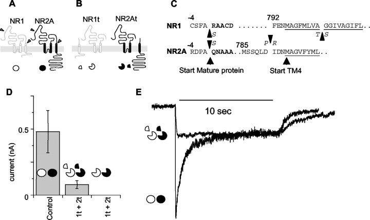
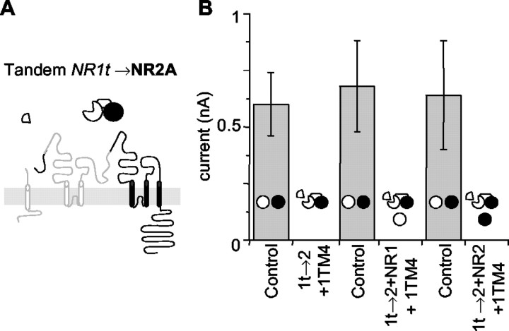
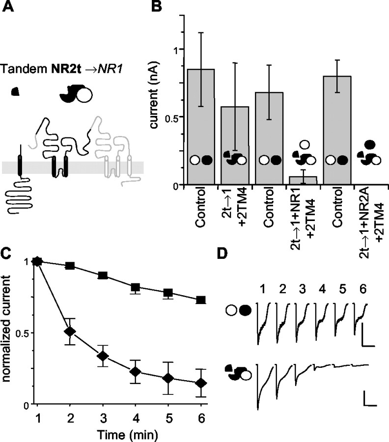
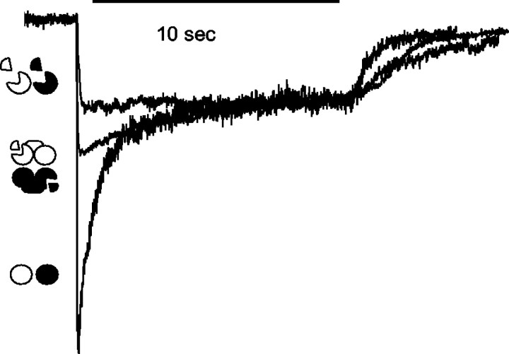
The subunits that compose eukaryotic glutamate ion channel receptors have three transmembrane domains (TMs) and terminate with intracellular tails that are important for controlling channel expression and localization. Truncation of NMDA receptor subunits before the final TM showed that this TM and intracellular tail region are necessary to form functional channels. However, it is shown here that these truncated subunits may be partially rescued by coexpressing the final TM and tail as a separate protein. The whole-cell currents so produced are somewhat lower than with full-length subunits, and they do not show the sag characteristic of currents from channels containing NR1 and NR2A subunits in the continued presence of an agonist. In addition, these truncated subunits were joined to full-length subunits to generate tandems. The functional expression of these tandems confirmed the tetrameric structure of NMDA receptors and also suggested that the subunits making up NMDA receptors are arranged as a dimer of dimers in the receptors with a 1-1-2-2 orientation of the subunits in the channel, and not in an alternating pattern of subunits around the pore. These results may redirect future studies into the mechanism of binding and gating in these receptors toward schemes including dimers, and may also be relevant to studies of glutamate receptor ion channels in general.


Figures

Fig. 1.
Fig. 1.
Generation and expression of truncated subunits.A, Schematic of subunit layout showing cut sites in NR1 and NR2a subunits. NR1 is white; NR2A isblack. The symbols underneath, awhite circle for NR1 and a black circlefor NR2A, are used throughout as a guide to the subunits included in each experiment. Full circles indicate full-length subunits. B, The putative membrane arrangement of the truncated subunits NR1t and NR2t and the arrangement of the TM4-tail pieces. Symbols are as in A, withwedges removed from the circles indicating truncation of the main subunits and quarter circle segments indicating TM4-tail peptides expressed as separate peptides linked to their own signal sequence. C, The exact amino acid sequences at the sites at which mutations were introduced to generate the cut sites. The single-letter code also indicates at what point conservative amino acid substitutions were introduced in the truncated subunits. Numbering is as in Anson et al. (1998), with the signal sequence negative from the start site of the mature protein. D, The average relative currents of cells expressing NR1 and NR2A subunits (Control) and truncated subunits with (middle) and without (right) the TM4-tail peptides coexpressed. Currents were elicited with 1 sec applications of 1 mm glutamate and 1 mm glycine 24–48 hr after transfection. Cells used in the chart were all recorded from transfections done at the same time on the same batch of cells. Currents were measured within 1 min of achieving whole-cell configuration. Symbols are as in A. Exact data are summarized in Table 1. 1t + 2t, Truncated NR1 plus truncated NR2 subunits. E, Representative traces from cells expressing control NR1 and NR2A channels and truncated NR1t and NR2t channels. Currents were elicited by a 10 sec pulse of 1 mm glutamate and 1 mm glycine in 2 mm CaCl2. The control, NR1 and NR2A trace (circles) shows the rapid sag characteristic of NR2A-containing NMDA channels. Under identical conditions, currents from cells expressing NR1t and NR2t show almost no sag (circles missing wedges). Currents in the figure are normalized at the 10 sec level.
Fig. 2.
Fig. 2.
1t→2 tandems fail to generate functional channels. A, The presumed membrane orientation of the NR1t→2 tandem. The TM4 tail for NR1, the subunit truncated to form the tandem, is included as a separate subunit. The symbol above indicates the order of subunits in the tandem, with thetruncated subunit missing a 90οsegment forming the start or head of the tandem (NR1, white). NR2, black. B, NR1t→2 tandems do not generate currents. Currents from NR1 and NR2A cells (Control) recorded on the same day as NR1t→2 tandems alone, or with different full-length subunits added, are shown. The NR1t→2 tandem gives no significant currents alone or with any combination of single subunits. Diagrams of tandems are included in the histograms to indicate which DNAs were introduced to the cells for each column. Data are given in Table2.
Fig. 3.
Fig. 3.
2t→1 tandems produce currents that run down quickly. A, The presumed membrane organization of the NR2t→1 tandem with its symbol above. The TM4 tail for NR2A, the subunit truncated to form this tandem, is included as a separate subunit. The symbol above is as in Figure2A, with black for NR2-derived portions and white for NR1. B, NR2t→1 tandems are able to produce unstable currents. As in Figure2B, cells expressing the NR2t→1 tandem were compared with cells transfected with NR1 and NR2A subunits in the same experiment. The 2t→1 alone was able to generate currents similar in amplitude to the control cells transfected on the same day. The addition of either NR1 or NR2A alone to the NR2t→1 tandem caused a dramatic drop in the currents produced by these cells.C, Currents derived from the NR2t→1 tandem rundown rapidly compared with control NR1 and NR2A cells. One second applications of 1 mm glutamate and 1 mm glycine were given at 1 min intervals starting within 1 min of obtaining the whole-cell configuration. All levels are normalized to the first application. The NR2t→1 tandem was supplemented with the NR2 TM4-tail segment in all cells. Squares, Controls;diamonds, 2t→1 only. D, Traces from representative cells showing rundown of NR2t→1 relative to control. The top traces are from a control cell, and thebottom traces are from a cell expressing NR2t→1. Calibration: 500 pA, 5 sec.
Fig. 4.
Fig. 4.
Predicted and aberrant arrangements of subunits included in tandem constructs. A, The theoretical arrangements of expressed single subunits and tandems and their ability to generate functional currents. Diagrams are as used in all figures;open areas represent NR1-derived elements; filled areas represent NR2A-derived elements. In all cases thesubunit missing a 90° segment is the start (the N terminal or head of a tandem), and the full circle is the end of the tandem. Arrangements shown in A are the organizations expected to be permitted by tandems without stretching or distorting the linker. B, Some of the possible ways that subunits in tandems may be able to violate the arrangements shown inA. The letters N and C are used to show the N and C terminals of the tandems forming a channel. Diagrams are as in A, except that gray-scale subunits indicate that the normally intracellular region of the tandem has flipped to the outside of the cell (flip in membrane). The side of the tandem used to form the pore has been chosen arbitrarily.C, Subunits within a single tandem may participate in the formation of two different channels. A chain of channels may be formed by subunits within a tandem participating in separate channels. The arrangement shown would allow a dimer of dimers organization of subunits within channels, although only the 2t→1 tandem is used.
Fig. 5.
Fig. 5.
Sustained currents are produced by mixtures of tandems allowing a 1-1-2-2 organization of subunits in receptors.A, Mixtures of pairs of tandems were able to generate sustained currents. Currents from cells expressing a mix of 1t→2 and 2t→1 (columns 2 and 3) or 1t→1 and 2t→2 (columns 4 and 5) are shown compared with currents from cells transfected with full-length NR1 and NR2A (Control). For clarity, the diagrams indicate the DNAs included as well as the axis labels. All currents resulted from 1 sec applications of 1 mm glutamate and 1 mm glycine applied within 1 min of obtaining a whole-cell configuration. In the presence of both of the TM4 tails (columns 2 and 4), both mixtures of tandems generated currents that were not significantly different from control cells. In the absence of the TM4 tails, the 1t→2 and 2t→1 mixture did elicit small currents in two of four cells. The 1t→1 and 2t→2 cells did not have currents when the TM4 tails were not coexpressed (0 of 10 cells; full data in Table 1). B, Currents generated from mixtures of tandems are more stable than those from 2t→1 alone. The rundown was measured as in Figure 3C.Squares, Control; diamonds, 2t→1 only;down triangles, 2t→1 with 1t→2 and both TM4 tails;up triangles, 1t→1 with 2t→2 with both TM4 tails. All currents are normalized to the first application. C, Representative traces from cells expressing mixtures of tandems. Thebottom two traces are from cells expressing both 2t→1 with 1t→2, showing different levels of rundown in this population. The top traces are from a representative cell expressing 1t→1 with 2t→2. Calibration: 250 pA, 5 sec for all traces.
Fig. 6.
Fig. 6.
Normalized currents from cells expressing 1t→1 with 2t→2 and both TM4 tails show an intermediate level of sag. Representative traces, as in Figure 1E, from 10 sec applications of 1 mm glutamate and 1 mmglycine show an intermediate level of sag in cells containing 1t→1 and 2t→2 with TM4 tails. Circles, Full-length NR1 and NR2A; circles missing wedges, NR1t and NR2t;connected symbols, 1t→1 with 2t→2. All currents are normalized to the 10 sec level.

References

    1. Anson LC, Chen PE, Wyllie DJ, Colquhoun D, Schoepfer R. Identification of amino acid residues of the NR2A subunit that control glutamate potency in recombinant NR1/NR2A NMDA receptors. J Neurosci. 1998;18:581–589. - PMC - PubMed
    1. Armstrong N, Gouaux E. Mechanisms for activation and antagonism of an AMPA-sensitive glutamate receptor: crystal structures of the GluR2 ligand binding core. Neuron. 2000;28:165–181. - PubMed
    1. Baumann SW, Baur R, Sigel E. Subunit arrangement of γ-aminobutyric acid type A receptors. J Biol Chem. 2001;276:36275–36280. - PubMed
    1. Behe P, Stern P, Wyllie DJ, Nassar M, Schoepfer R, Colquhoun D. Determination of NMDA NR1 subunit copy number in recombinant NMDA receptors. Proc R Soc Lond B Biol Sci. 1995;262:205–213. - PubMed
    1. Bigge CF. Ionotropic glutamate receptors. Curr Opin Chem Biol. 1999;3:441–447. - PubMed

Publication types

LinkOut - more resources