. 2003 Feb 15;23(4):1169-78.
doi: 10.1523/JNEUROSCI.23-04-01169.2003.

Neuronal hyperpolarization-activated pacemaker channels drive neuropathic pain

Affiliations

Neuronal hyperpolarization-activated pacemaker channels drive neuropathic pain

Sandra R Chaplan et al. J Neurosci. .

Abstract

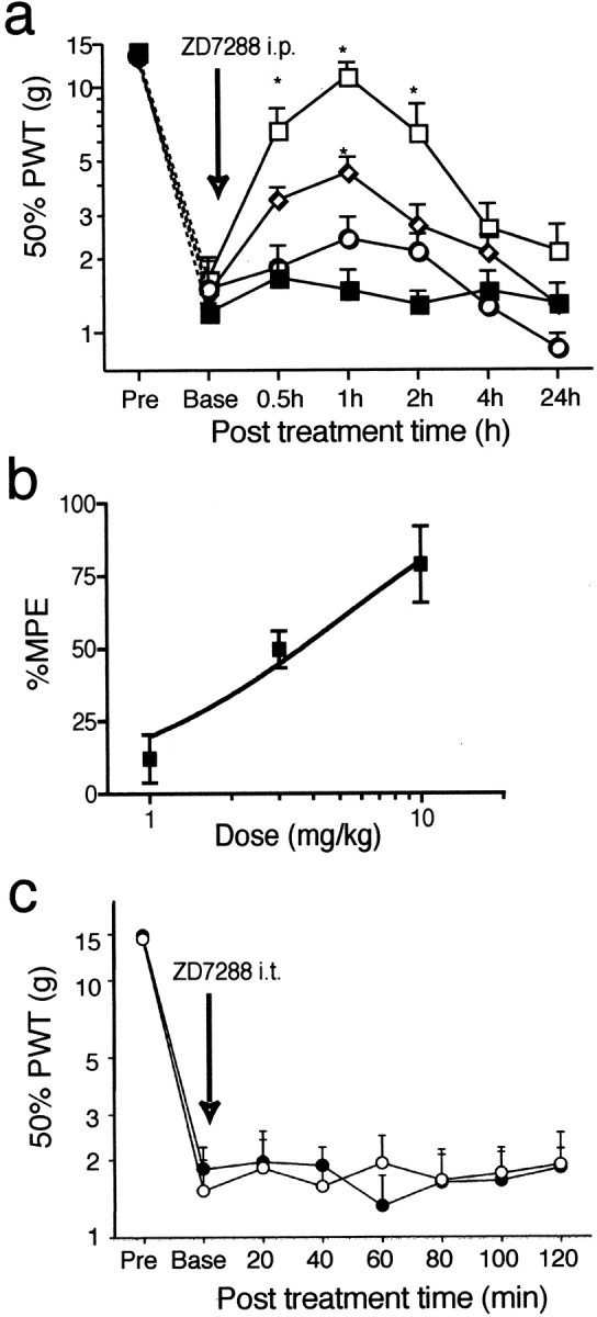
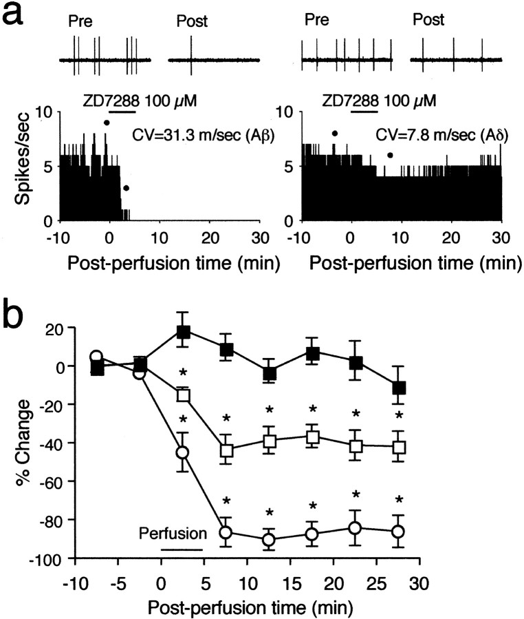
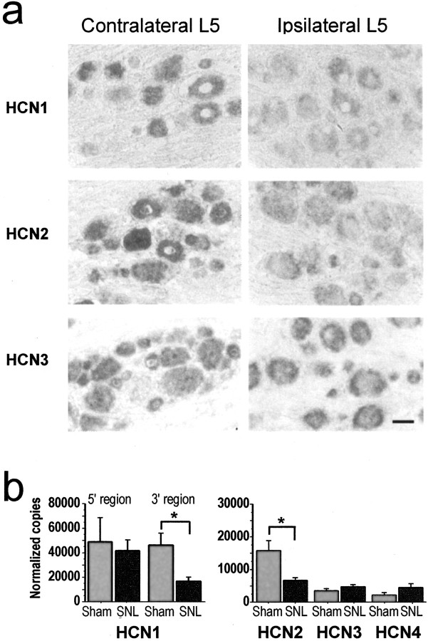
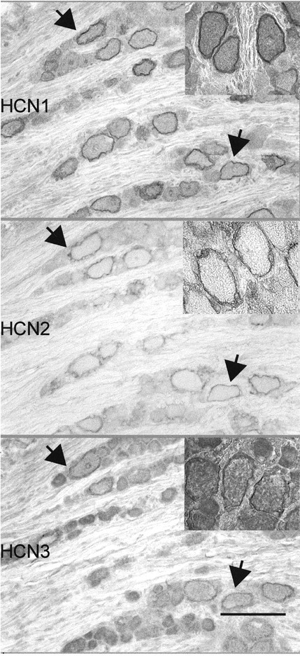
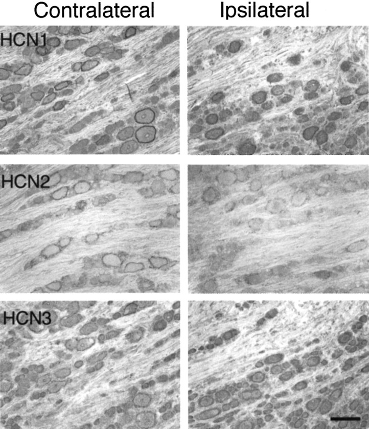
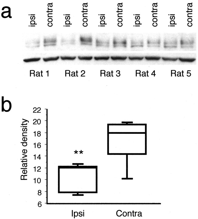
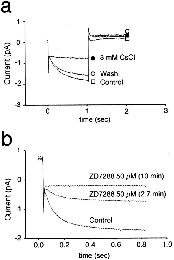
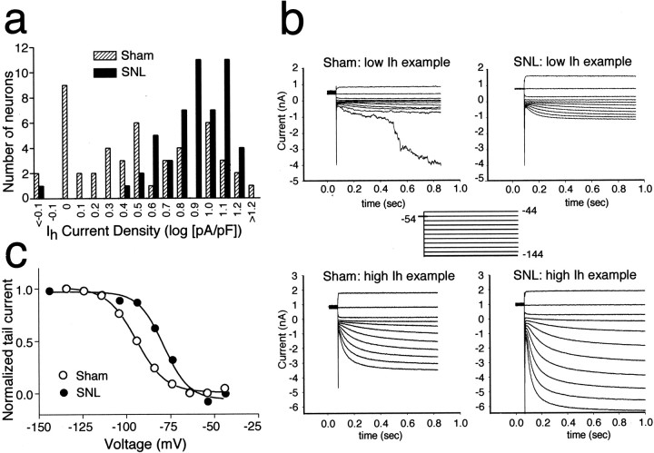
Neuropathic pain is a common and often incapacitating clinical problem for which little useful therapy is presently available. Painful peripheral neuropathies can have many etiologies, among which are trauma, viral infections, exposure to radiation or chemotherapy, and metabolic or autoimmune diseases. Sufferers generally experience both pain at rest and exaggerated, painful sensitivity to light touch. Spontaneous firing of injured nerves is believed to play a critical role in the induction and maintenance of neuropathic pain syndromes. Using a well characterized nerve ligation model in the rat, we demonstrate that hyperpolarization-activated, cyclic nucleotide-modulated (HCN) "pacemaker" channels play a previously unrecognized role in both touch-related pain and spontaneous neuronal discharge originating in the damaged dorsal root ganglion. HCN channels, particularly HCN1, are abundantly expressed in rat primary afferent somata. Nerve injury markedly increases pacemaker currents in large-diameter dorsal root ganglion neurons and results in pacemaker-driven spontaneous action potentials in the ligated nerve. Pharmacological blockade of HCN activity using the specific inhibitor ZD7288 reverses abnormal hypersensitivity to light touch and decreases the firing frequency of ectopic discharges originating in Abeta and Adelta fibers by 90 and 40%, respectively, without conduction blockade. These findings suggest novel insights into the molecular basis of pain and the possibility of new, specific, effective pharmacological therapies.


Figures

Fig. 1.
Fig. 1.
The HCN antagonist ZD7288 reverses neuropathic pain behavior. a, One week after SNL, rats received equivalent volumes of intraperitoneal saline (▪), 1 mg/kg ZD7288 (○), 3 mg/kg ZD7288 (⋄), or 10 mg/kg ZD7288 (■) after baseline assessment of tactile allodynia with von Frey hairs. The redacted time course of allodynia suppression is illustrated:y-axis = 50% paw withdrawal threshold (PWT) (normal = 15 gm; allodynia = lower threshold values). At 10 mg/kg, near complete suppression of allodynia is seen. *p < 0.05, one-way ANOVA with Dunnett's multiple comparisons; n = 7–8 per group. b, The dose–response curve for ZD7288 froma: y-axis: percentage of maximum possible effect (MPE) (15 gm threshold = 100% allodynia suppression; no change from pre-drug baseline = 0%). The ED50 for allodynia suppression is ∼3 mg/kg.c, Lumbar intrathecal administration of ZD7288, 50 μg (●), had no effect on SNL-related tactile allodynia at any time point, compared with saline (○), over a 2 hr observation period:y-axis: 50% paw withdrawal threshold (PWT).
Fig. 2.
Fig. 2.
Inhibition of HCN channels suppresses ectopic neuronal discharge without conduction blockade. a, Histogram (y-axis = spikes/sec) for single fiber ex vivo recording from typical fibers before and after application of 100 μm ZD7288. The left panel illustrates an Aβ fiber showing complete suppression of ectopic firing 3–4 min after drug application. Dotsindicate the sources of 1 sec top panels showing spike patterns before (Pre) and after (Post) drug application on a faster time base. The right panelsimilarly illustrates single-fiber recording from a representative Aδ fiber showing attenuation of firing after ZD7288 application.b, Time course of percentage change in firing after ZD7288 application: mean ± SEM for seven to eight fibers per group: filled squares = ACSF control (Aβ and Aδ fibers combined); open squares = Aδ fibers;open circles = Aβ fibers. *p< 0.05, one-way ANOVA, Dunnett's multiple comparisons.
Fig. 3.
Fig. 3.
Distribution and quantification of HCN mRNAs after nerve injury. a, In situ hybridization: comparisons of co-embedded contralateral (uninjured) and ipsilateral (injured) L5 DRGs hybridized with antisense probes for HCN1, HCN2, or HCN3 show a visible reduction in both HCN1 and HCN2 message. No hybridization was seen using sense probes. Scale bar, 50 μm.b, Relative quantification of HCN mRNA transcripts in sham versus SNL L5/6 DRGs (normalized copy number per 2 DRGs) by real-time fluorescent quantitative PCR. In the SNL samples, there is a significant reduction in a 3′ but not in a 5′ HCN1 amplicon compared with controls, a significant reduction in HCN2, and no change in HCN3 or HCN4 (*p < 0.02, unpaired ttest; n = 8 each group).
Fig. 4.
Fig. 4.
Antibody validation. a, Strong immunoreactivity to a stable HEK293–recombinant human HCN1 cell line is seen using anti-HCN1 antibody (left,HEK+HCN1). Immunoreactivity is absent in the parent (untransfected) cell line (HEK−) using the same antibody. b, Western blot analysis shows no anti-HCN1 antibody recognition of extracts from untransfected (HEK−) cells and a specific double band pattern in HEK+HCN1 cell extracts (left). Right, Single bands are identified in DRG and stable HEK-tsA201–recombinant human HCN3 (HEK+HCN3) cell line extracts using anti-HCN3 antibody, with absence of staining of HEK− cells.
Fig. 5.
Fig. 5.
HCN1, HCN2, and HCN3 colocalize in neuronal membranes in DRG. Immunohistochemical visualization of colocalization of HCN1-, HCN2-, and HCN3-IR in neuronal membranes, in adjacent (serial) 10 μm sections from normal DRG, is shown. Colocalization can be seen in numerous neuronal profiles. Immunoreactive profiles are predominantly large neurons. Arrows indicate two of the neurons best seen in all sections. Scale bar, 100 μm.
Fig. 6.
Fig. 6.
HCN1 and HCN2 membrane IRs are reduced in DRG after nerve injury. Immunohistochemical visualization of HCN1-, HCN2-, and HCN3-IR in simultaneously processed contralateral (uninjured) and ipsilateral (injured) L5 DRGs is shown. Note decreases in HCN1- and HCN2-IR in injured DRG cell membranes. Scale bar, 100 μm.
Fig. 7.
Fig. 7.
Total HCN1-IR is decreased in DRG after nerve injury. a, Western blot analysis was performed on DRGs from five rats 1 week after SNL. Anti-HCN1 antibody was used to compare immunoreactivity in protein extracts from ipsilateral (injured) and contralateral (uninjured) DRGs (top bands). Anti-α-tubulin antibody was used to correct for loading errors (bottom bands). b, Densitometric analysis revealed a significant decrease in HCN1-IR in ipsilateral DRGs compared with contralateral DRGs after normalization to α-tubulin IR. Thebox plot depicts the median and interquartile ranges;y-axis = arbitrary density units. **p < 0.02, t test.
Fig. 8.
Fig. 8.
Pharmacological blockade ofIh in dissociated DRG neurons. The pharmacology of hyperpolarization-induced inward currents in large SNL neurons was consistent with that of Ih. Currents were blocked by bath application of 3 mm CsCl (a) and 50 μm ZD7288 (b). a,Ih was elicited by voltage steps from −54 to −114 mV (time 0–1 sec) followed by a step to −54 mV applied every 15 sec. Initially a large slowly developing inward (downward deflection) current was observed after the initial capacitative transient (Control, ■). After exposure to 3 mm CsCl, the inward current was blocked (3 mm CsCl, ●), leaving only a CsCl-insensitive leak current (leak subtraction was not performed in these recordings). The inhibition by CsCl was reversed after washout (Wash, ○). b,Ih was elicited by voltage steps from −54 to −124 mV applied every 15 sec. Bath application of 50 μm ZD7288 caused a slow onset block ofIh that achieved steady state by 10 min. The effect of ZD7288 was not reversible over 30 min.
Fig. 9.
Fig. 9.
Nerve injury increasesIh density and depolarizesIh activation threshold.Ih expression in large-diameter rat DRG neurons was increased after ligature of the L5 peripheral nerve (SNL). a, The distribution ofIh peak current at the end of the test pulse was normalized to cell size (as measured by cell capacitance).Ih was determined in both control (hatched bars) and SNL L5 neurons (solid bars) at a step to −114 mV. The distribution was skewed toward higher Ih densities in SNL-operated compared with controls. b, Examples of families of hyperpolarization-activated currents elicited in sham-injured and injured DRG neurons elicited by the voltage protocol shown in theinset. Top of panelillustrates neurons with low-density Ih as seen in either sham or injured DRGs; bottom ofpanel illustrates neurons in both treatment categories with high-density Ih. The number of neurons with abundant Ih was greatly increased after nerve injury. Currents were elicited by −10 mV incremental voltage steps between −44 and −144 mV (inset).c, Voltage dependence of activation derived from tail current analysis in control and SNL neurons. In SNL neurons, the dependence was shifted to more depolarized potentials (+10 mV) without a significant change in the slope.

Similar articles

Cited by

References

    1. Abdi S, Lee DH, Park SK, Chung JM. Lack of pre-emptive analgesic effects of local anaesthetics on neuropathic pain. Br J Anaesth. 2000;85:620–623. - PubMed
    1. Abdulla FA, Smith PA. Axotomy- and autotomy-induced changes in Ca2+ and K+ channel currents of rat dorsal root ganglion neurons. J Neurophysiol. 2001;85:644–658. - PubMed
    1. Amir R, Michaelis M, Devor M. Burst discharge in primary sensory neurons: triggered by subthreshold oscillations, maintained by depolarizing afterpotentials. J Neurosci. 2002;22:1187–1198. - PMC - PubMed
    1. Beaumont V, Zucker RS. Enhancement of synaptic transmission by cyclic AMP modulation of presynaptic Ih channels. Nat Neurosci. 2000;3:133–141. - PubMed
    1. BoSmith RE, Briggs I, Sturgess NC. Inhibitory actions of ZENECA ZD7288 on whole-cell hyperpolarization activated inward current (If) in guinea-pig dissociated sinoatrial node cells. Br J Pharmacol. 1993;110:343–349. - PMC - PubMed

Publication types

MeSH terms

LinkOut - more resources