. 2003 Feb 15;23(4):1340-50.
doi: 10.1523/JNEUROSCI.23-04-01340.2003.

Functional role of C-terminal cytoplasmic tail of rat vanilloid receptor 1

Affiliations

Functional role of C-terminal cytoplasmic tail of rat vanilloid receptor 1

Viktorie Vlachová et al. J Neurosci. .

Abstract

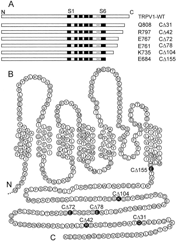
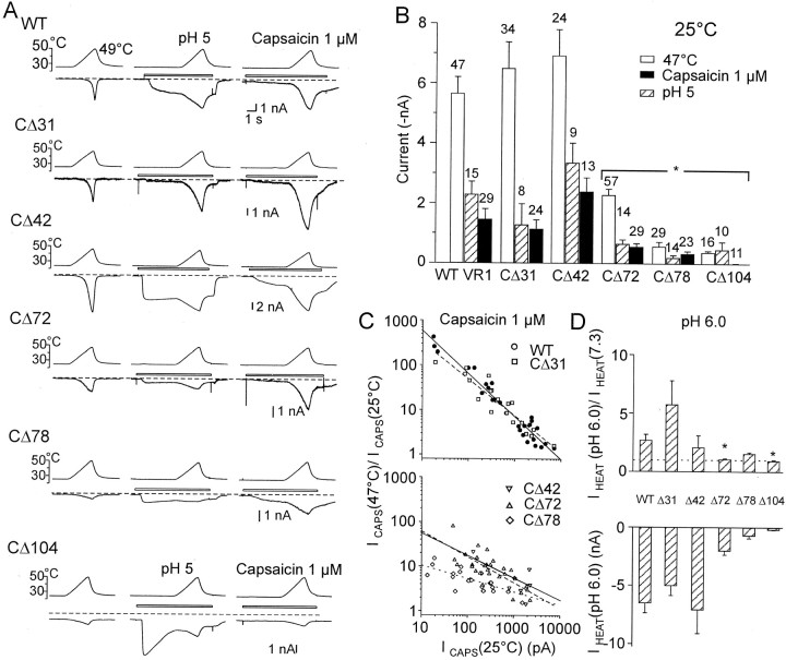
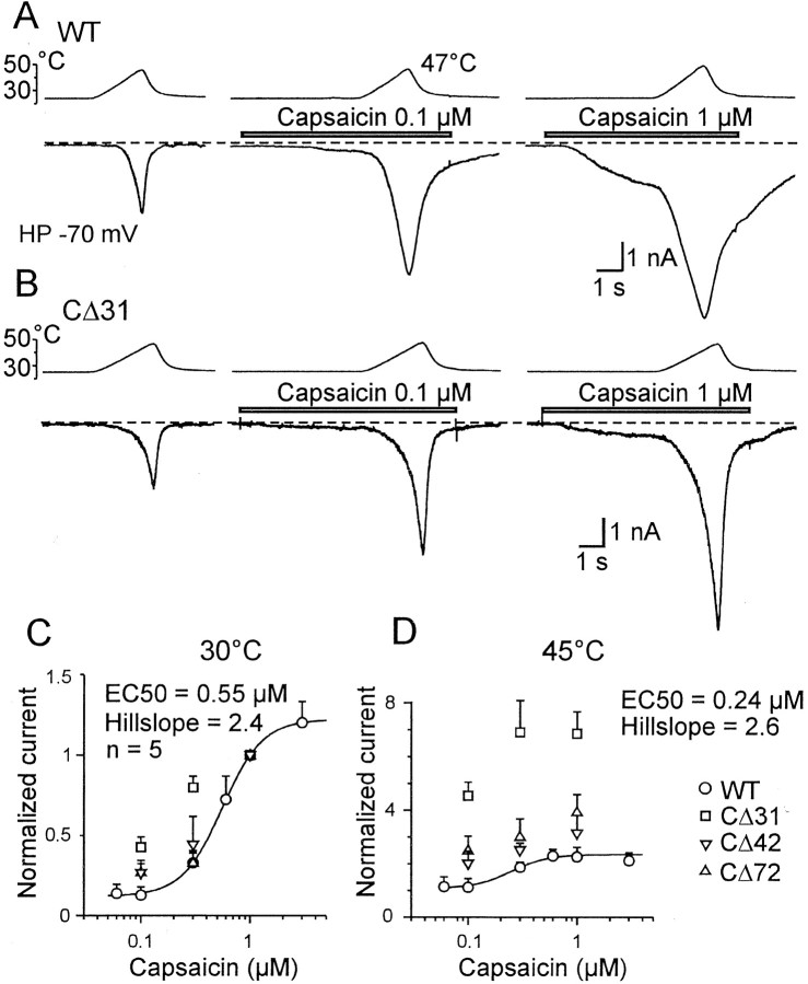
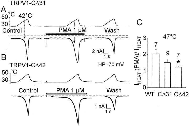
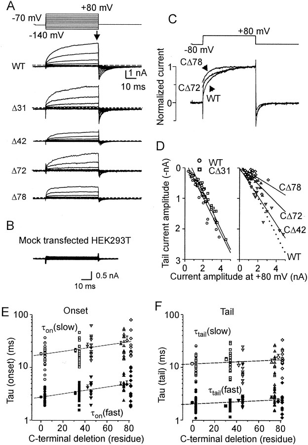
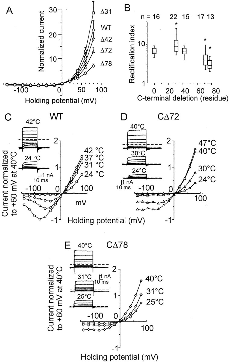
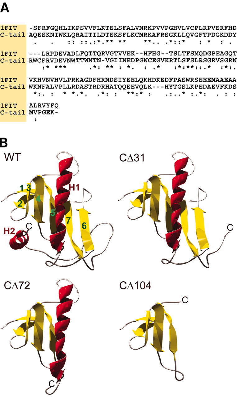
The vanilloid receptor [transient receptor potential (TRP)V1, also known as VR1] is a member of the TRP channel family. These receptors share a significant sequence homology, a similar predicted structure with six transmembrane-spanning domains (S1-S6), a pore-forming region between S5 and S6, and the cytoplasmically oriented C- and N-terminal regions. Although structural/functional studies have identified some of the key amino acids influencing the gating of the TRPV1 ion channel, the possible contributions of terminal regions to vanilloid receptor function remain elusive. In the present study, C-terminal truncations of rat TRPV1 have been constructed to characterize the contribution of the cytoplasmic C-terminal region to TRPV1 function and to delineate the minimum amount of C tail necessary to form a functional channel. The truncation of 31 residues was sufficient to induce changes in functional properties of TRPV1 channel. More pronounced effects of C-terminal truncation were seen in mutants lacking the final 72 aa. These changes were characterized by a decline of capsaicin-, pH-, and heat-sensitivity; progressive reduction of the activation thermal threshold (from 41.5 to 28.6 degrees C); and slowing of the activation rate of heat-evoked membrane currents (Q10 from 25.6 to 4.7). The voltage-induced currents of the truncated mutants exhibited a slower onset, markedly reduced outward rectification, and significantly smaller peak tail current amplitudes. Truncation of the entire TRPV1 C-terminal domain (155 residues) resulted in a nonfunctional channel. These results indicate that the cytoplasmic COOH-terminal domain strongly influences the TRPV1 channel activity, and that the distal half of this structural domain confers specific thermal sensitivity.


Figures

Fig. 1.
Fig. 1.
C-terminal truncations of vanilloid receptor TRPV1. A, Schematic representation of TRPV1 C-terminal deletions showing the six transmembrane-spanning domains, S1–S6, and a pore-forming region (hatched bars).B, Putative membrane topology and amino acids (in circles) of the C-terminal half of the rat TRPV1 receptor with indicated locations of the amino acids at which the stop codon has been introduced. CΔ31, CΔ42, CΔ72, CΔ78, CΔ104, and CΔ155 correspond to the mutated Q808, R797, E767, E761, K735, and E684, respectively.
Fig. 2.
Fig. 2.
Whole-cell membrane currents induced by heat, protons, and capsaicin in wild-type TRPV1 and the C-terminal truncated mutants expressed in HEK293T cells. A, Heat-evoked currents (IHEAT) were induced by a 3 sec ramp of temperature increase to 49°C in standard extracellular solution (left), acidic pH (pH 5;middle), and capsaicin (1 μm;right). The cells clamped at −70 mV were superfused for the time indicated by the open bars shown above the records. Dashed lines indicate a zero membrane current.B, Quantitative analysis was performed by first stimulating cells by heat (from 25 to 49°C) in the extracellular solution, followed by a 30 sec washout, and then by stimulating them by heat in the presence of pH 5 or capsaicin. Mean ± SEM responses evoked by heat, capsaicin, and low pH measured at room temperature, 25°C, are shown (p < 0.05; indicated with an asterisk). The numbersabove each bar indicate the number of cells measured.C, Relative increase in capsaicin-induced peak currents at 47°C versus steady-state capsaicin current amplitudes measured at 25°C are plotted for individual cells transfected with wild-type and CΔ31 (top) and CΔ42, CΔ72, and CΔ78 (bottom). D, Analysis of proton-induced potentiation of heat-evoked currents. Mean ratios of peak currents induced by heat (47°C) at pH 6 to those measured at pH 7.3 are shown at the top. Average amplitudes of heat-induced currents recorded at pH 6 are shown at the bottom. Data represent means ± SEM from 8 to 21 cells.
Fig. 3.
Fig. 3.
The effects of TRPV1 C-terminal truncation on receptor capsaicin efficacy and potency. A, Representative traces of whole-cell currents in WT TRPV1 activated by a heat ramp from 24 to 47°C in normal extracellular solution (left), 0.1 μm capsaicin (middle), and 1 μm capsaicin (right). The holding potential (HP) is −70 mV. B, The HEK293T cell transiently transfected with mutant CΔ31 was exposed to heat ramps from 24 to 47°C in normal extracellular solution (left), 0.1 μm capsaicin (middle), and 1 μm capsaicin (right). The cells clamped at −70 mV were superfused for the time indicated by the open bars shown above the records. Dashed linesindicate a zero membrane current. C, The currents were normalized to the current produced by the application of 1 μm capsaicin at 30°C and the mutants CΔ31, CΔ42, and CΔ72 were tested at agonist concentrations of 0.1, 0.3, and 1 μm. At 30°C, capsaicin dose–response curves for wild-type receptor exhibited the average half-maximal concentration 0.55 μm and Hill slope 2.4 (n = 5). The Hill equation was used for fitting control wild-type data (solid line). Before averaging across cells, data from each recording were normalized to the current induced by 1 μm capsaicin measured at 30°C. Data for each construct are shown as the means ± SEM for four independent measurements. D, At 45°C, capsaicin dose–response curves for wild-type receptor exhibited the average half-maximal concentration 0.24 μm and Hill slope 2.6 (n = 5). Data from each recording were normalized to the current induced by 1 μm capsaicin measured at 30°C. C-terminal truncation had the most pronounced effects on both agonist efficacy and agonist potency in mutant CΔ31.
Fig. 4.
Fig. 4.
Sensitivity to heat in C-terminally truncated mutants. A, Representative heat-induced responses recorded from HEK293T cells transiently transfected with wild-type TRPV1, CΔ31, CΔ42, CΔ72, CΔ78, and CΔ104 mutants. Heat-evoked currents were induced by a 3 sec temperature ramp (24–49°C) in control extracellular solution. Dashed lines indicate a zero membrane current. HP, Holding potential.B, Temperature response profiles of the heat-induced currents shown in A. The threshold forIHEAT was 43.8, 38.2, and 28.9°C for wild-type, CΔ31, and CΔ42, respectively (left). In CΔ104 mutant, a pronounced resting membrane current was observed at room temperature (right). C, Arrhenius plot in which the current (y-axis, log scale) was plotted against the reciprocal of the absolute temperature (x-axis). The temperature coefficient,Q10, was estimated for each mutant by linear regression from the slope of the Arrhenius plot in the temperature range indicated by respective symbols.Q10 was 104.1, 22.1, 20.5, 5.9, 1.8, and 1.2 for wild-type TRPV1, CΔ31, CΔ42, CΔ72, CΔ78, and CΔ104 mutants, respectively. D, Thermal threshold values plotted versus the number of residues deleted from the C-terminal tail of TRPV1 receptor. Sequential deletions of 31, 42, and 72 residues from the C-terminal end resulted in a significant shift of the thermal threshold to 38.6 ± 0.7°C (SEM; n = 22), 32.5 ± 0.5°C (n = 21), and 28.6 ± 0.5 (n = 50), respectively. Deletions of 78 and 104 residues from the C-terminal end caused a shift of the TRPV1 thermal threshold toward temperatures close to that in the bath.E, Q10 distribution in wild-type exhibits a median value of 25.6 (n = 26); the histogram shows that this distribution is similar to the distribution of Q10 in the CΔ31 mutant (also see Table 1). F, The medianQ10 plotted versus the number of residues deleted from the C-terminal tail of TRPV1 receptor. Theends of the boxes define the 25th and 75th percentiles, with a line at the median; error bars define the 10th and 90th percentiles. The asterisk indicates statistically significant difference from the wild type (p < 0.05).
Fig. 5.
Fig. 5.
Effects of PMA on heat-evoked current in CΔ31 mutant and the construct lacking the Ser residue at position 800, CΔ42. A, Heat-evoked currents were induced by ramps of increasing temperature from 25 to 47°C (8°C/sec) in control extracellular solution (Control) and in the presence of 1 μm PMA followed by a wash-off with control (Wash). The cells clamped at −70 mV were superfused for the time indicated by the open bars shown above the records. Dashed lines indicate a zero membrane current. B, Membrane currents recorded from TRPV1–CΔ42-transfected HEK293T cells. C, PMA-induced potentiation of the heat-evoked currents at 47°C in CΔ42 compared with the wild-type and CΔ31 mutant (p < 0.05; indicated with an asterisk). Data represent means ± SEM; the numbers above eachbar indicate the number of cells measured.
Fig. 6.
Fig. 6.
Voltage-activated currents in wild-type TRPV1 and C-terminal truncated mutants. A, Representative whole-cell membrane currents induced by a sequentially applied series of 50 msec voltage steps ranging from −140 to +80 mV, in +20 mV increments. The currents were measured at the end of each pulse (arrow). B, The voltage protocol was completely ineffective in a mock-transfected HEK293T cell.C, Superimposed example responses induced by a depolarizing step from −80 to +80 mV in wild-type and the CΔ72 and CΔ78 mutants. D, The ratio between the outward current at +80 mV and the tail current amplitude in CΔ31 and CΔ42 is not statistically different from wild-type TRPV1 (Spearman correlation; r = −0.98 and −0.95 vs −0.96 for wild-type); on the contrary, it is markedly lower in CΔ72 and CΔ78 mutants (−0.86 and −0.68, respectively). E, Kinetic analysis of the outward currents recorded from 36, 24, 12, 17, and 14 cells transfected with wild-type and CΔ31, CΔ42, CΔ72, and CΔ78 mutants, respectively. The onset currents induced by the depolarizing step from −80 to +80 mV were best fitted by the sum of two exponential functions with the slow component [τon(slow);open symbols], and the fast component [τon(fast);filled symbols]. Averages represent means ± SEM. Linear regression analysis of the data yielded slopes of 0.0031 (r = 0.94) and 0.0033 (r = 0.93) for the slow and the fast components, respectively.F, The tail currents were best fitted by the sum of two exponential functions: f (t) = Afastexp[−ttail (fast)] +Aslowexp[−ttail (slow)]. The regression lines (dashed) were shallow, possessing slopes of 0.0011 (r = 0.27) and 0.0011 (r = 0.33) for the slow and the fast components, respectively.
Fig. 7.
Fig. 7.
Outward currents in HEK293T cells transiently transfected with wild-type TRPV1 and C-terminal truncated mutants.A, Current–voltage relationship for wild-type and CΔ31, CΔ42, CΔ72, and CΔ78 mutants. Data were normalized to the value of the current at −40 mV. B, For CΔ31, RI was significantly higher than that for wild-type, whereas significantly less RI was observed for CΔ72 and CΔ78 mutants (Table 2). Theends of the boxes define the 25th and 75th percentiles, with a line at the median; error bars define the 10th and 90th percentiles. The asterisks indicate significant differences from wild type (p < 0.05).C, The effect of temperature on the outward current rectification. Representative voltage-gated responses, recorded at 24°C (inset, bottom record) and at 42°C (inset, top record). Dashed lines indicate a zero membrane current in all records in this figure. Data were normalized to the value of the current measured at +60 mV and at 42°C. D, Effect of temperature on outward current rectification properties in CΔ72 mutant. Representative recordings of voltage-evoked currents at 24, 30, and 40°C (inset). E, Note a considerable steady-state inward current induced at 31°C, a temperature at which the mutant CΔ78 is thermally activated.
Fig. 8.
Fig. 8.
Surface expression of C-terminally truncated TRPV1 construct in transiently transfected HEK293T cells. A, Confocal microscope image of wild-type transfected HEK293T cells. Permeabilized cells were immunohistochemically labeled using a polyclonal antibody against the first 21 aa residues of the rat TRPV1 N-terminal end. Cells were labeled with FITC-conjugated donkey anti-rabbit IgG secondary antibody and visualized with standard FITC filters. B, Four HEK293T cells transiently transfected with truncated construct lacking the entire C-terminal end CΔ155. Scale bar, 5 μm.
Fig. 9.
Fig. 9.
Molecular modeling of the TRPV1 C terminal.A, Sequence alignment of the cytoplasmic C terminal of TRPV1 from A690 to K838 and the template protein FHIT. Identical and similar amino acids of the stronger groups are indicated with anasterisk and colon, respectively. These amino acids should conserve the structure with a probability of >95%.Dots indicate similar amino acids of the lower groups that should conserve the structure with a lower probability.B, Predicted structure of the complete C terminal of TRPV1 and the truncated mutants. WT, Ribbon diagram of the wild-type C terminus (residues A690–K838). Homology modeling predicts two α-helices (H1, H2) and seven β-strands (1–7). Antiparallel strands 1 and 2 form a β-hairpin, and strands 3–7 form a five-stranded antiparallel sheet.CΔ31, This mutant (residues A690–T807) lacks the α-helix H2. CΔ72, In this construct (A690–C766), secondary structural elements H2 and β-strands 6 and 7 are missing.CΔ104, The predicted structure of the truncated construct (A690–G734) consists only of β-strands 1–5.

References

    1. Aydar E, Palmer C. Functional characterization of the C-terminus of the human ether-a-go-go-related gene K+ channel (HERG). J Physiol (Lond) 2001;534:1–14. - PMC - PubMed
    1. Bentley GN, Brooks MA, O'Neill CA, Findlay JB. Determinants of potassium channel assembly localised within the cytoplasmic C-terminal domain of Kv2.1. Biochim Biophys Acta. 1999;1418:176–184. - PubMed
    1. Bruening-Wright A, Schumacher MA, Adelman JP, Maylie J. Localization of the activation gate for small conductance Ca2+-activated K+ channels. J Neurosci. 2002;22:6499–6506. - PMC - PubMed
    1. Caterina MJ, Schumacher MA, Tominaga M, Rosen TA, Levine JD, Julius D. The capsaicin receptor: a heat-activated ion channel in the pain pathway. Nature. 1997;389:816–824. - PubMed
    1. Caterina MJ, Rosen TA, Tominaga M, Brake AJ, Julius D. A capsaicin-receptor homologue with a high threshold for noxious heat. Nature. 1999;398:436–441. - PubMed

Publication types

MeSH terms

LinkOut - more resources