. 2003 Feb 15;23(4):1372-82.
doi: 10.1523/JNEUROSCI.23-04-01372.2003.

Honeycomb-like mosaic at the border of layers 1 and 2 in the cerebral cortex

Affiliations

Honeycomb-like mosaic at the border of layers 1 and 2 in the cerebral cortex

Noritaka Ichinohe et al. J Neurosci. .

Abstract

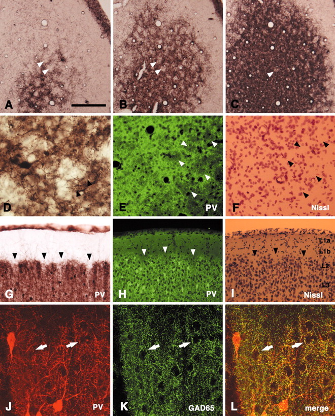
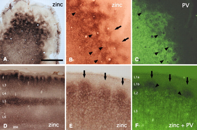
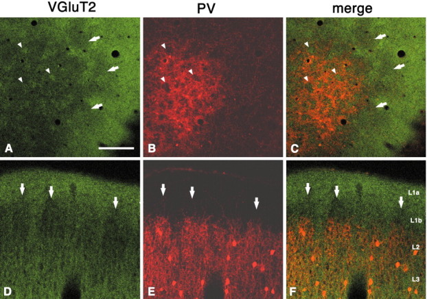
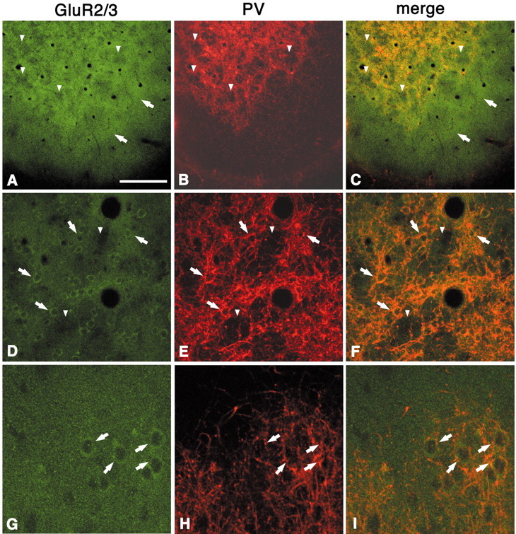
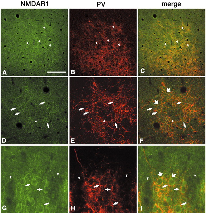
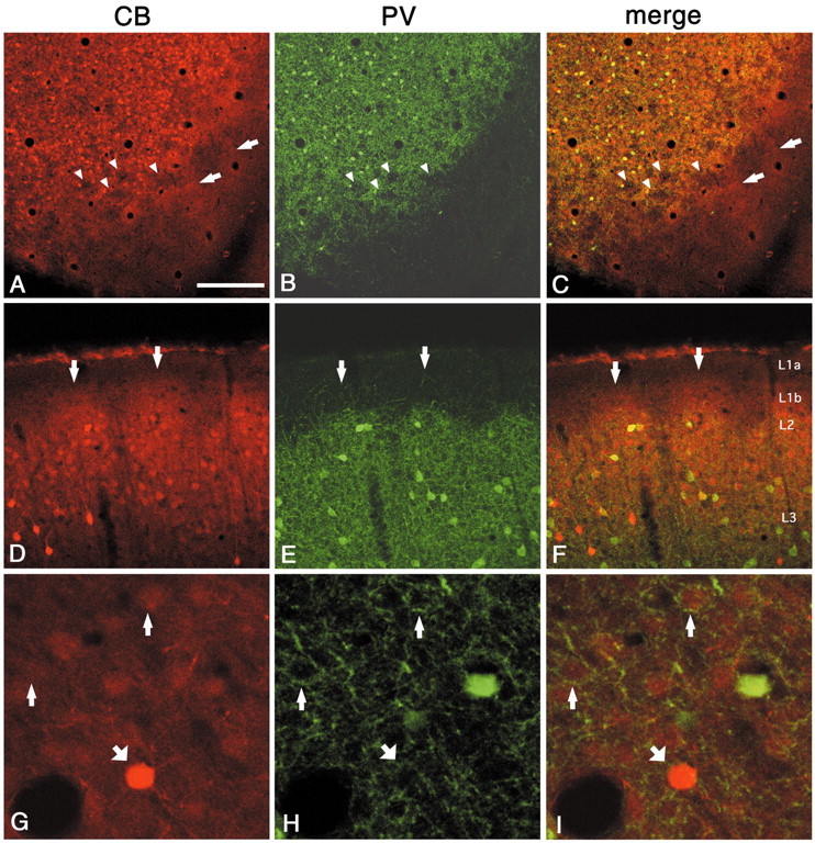
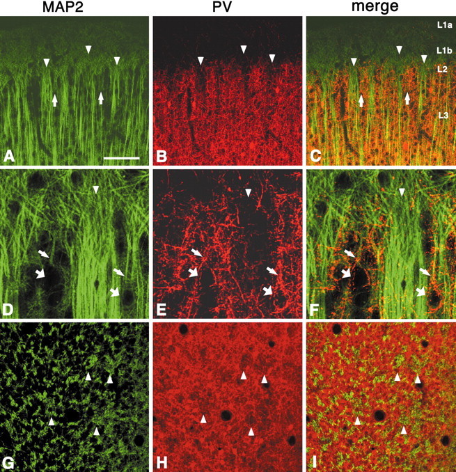
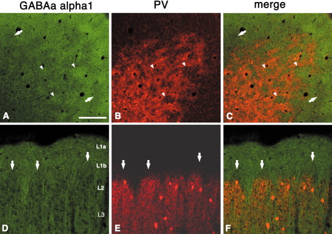
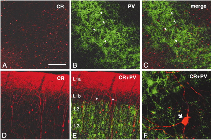
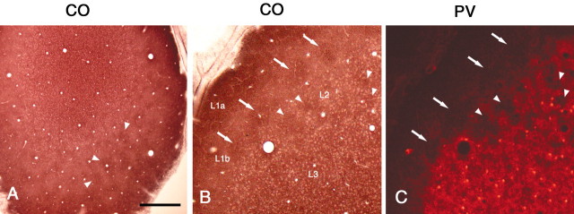
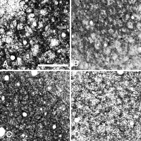
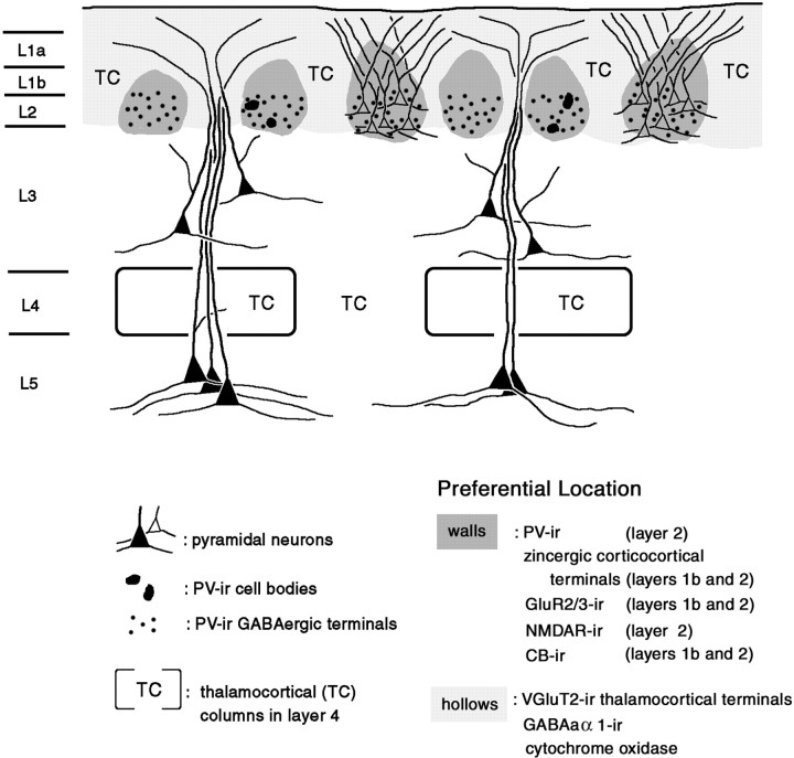
In this report, we present evidence of a small-scale modularity (<100 microm) at the border of layers 1 and 2 in neocortical areas. The modularity is best seen in tangential sections, with double-labeling immunohistochemistry to reveal overlapping or complementary relationships of different markers. The pattern is overall like a reticulum or mosaic but is described as a "honeycomb," in which the walls and hollows are composed of distinct afferent and dendritic systems. We demonstrate the main components of the honeycomb in rat visual cortex. These are as follows: (1) zinc-enriched, corticocortical terminations in the walls, and in the hollows, thalamocortical terminations (labeled by antibody against vesicular glutamate transporter 2 and by cytochrome oxidase); (2) parvalbumin-dense neuropil in the walls that partly colocalizes with elevated levels of glutamate receptors 2/3, NMDAR receptor 1, and calbindin; and (3) dendritic subpopulations preferentially situated within the walls (dendrites of layer 2 neurons) or hollows (dendrites of deeper neurons in layers 3 and 5). Because the micromodularity is restricted to layers 2 and 1b, without extending into layer 3, this may be another indication of a laminar-specific substructure at different spatial scales within cortical columns. The suggestion is that corticocortical and thalamocortical terminations constitute parallel circuits at the level of layer 2, where they are segregated in association with distinct dendritic systems. Results from parvalbumin staining show that the honeycomb mosaic is not limited to rat visual cortex but can be recognized at the layer 1-2 border in other areas and species.


Figures

Fig. 1.
Fig. 1.
PV immunoreactivity shows a honeycomb pattern in superficial layers in the rat V1. A–C, Serial tangential sections stained by immunoperoxidase method for PV. PV-ir walls surround open hollows (arrowheads).D, Higher magnification. Arrowheads point to PV-ir terminal-like puncta. E, F, Tangential section. Double staining for PV (immunofluorescence,E) and Nissl substance (F) shows that the hollows tend to contain fewer cells than do the walls.Arrowheads point to corresponding spaces.G, Coronal section stained by immunoperoxidase method for PV. The hollows are less conspicuous but can be detected as a series of notches (arrowheads). H, I,Coronal section. Double staining for PV (H) and Nissl substance (I) confirms that the honeycomb is located at the level of layer 2. Arrowheads point to corresponding hollows. J–L, Coronal section. Double-immunofluorescent staining for PV and GAD65 demonstrates colocalization of these two markers, as expected if the small PV-ir particles were GABAergic terminals. Double-labeled structures frequently made basket-like terminations on immunonegative somata (arrows). Scale bar (shown in A):AC, 200 μm; D, 80 μm; EI, 100 μm;JL, 25 μm.
Fig. 2.
Fig. 2.
Zinc-enriched terminals are located in the walls.A, Tangential section. Zinc walls surround open hollows.B, C, Tangential section. Double labeling for zinc and PV indicates that the zinc pattern closely colocalizes with the PV walls in layer 2; however, the zinc walls extend into layer 1b (arrows). Arrowheads point to corresponding hollows in both markers. D, Zinc staining, in coronal section, shows a conspicuous modularity at the border of layers 1 and 2. Lateral is to the left.E, F, Double labeling for zinc and PV confirms that zinc walls (arrows) extend into layer 1b, higher than PV walls (arrowheads). WM, White matter. Scale bar (shown in A): A,D, 500 μm; B, C,E, F, 100 μm.
Fig. 3.
Fig. 3.
Immunohistochemistry for VGluT2 shows dense uniform staining in layer 1a but a discontinuous periodic pattern in layers 1b and 2 (A–C, tangential section;D–F, coronal section). Double labeling for VGluT2 and PV shows that VGluT2-ir dense regions in layer 2 are situated within the PV hollows. Arrowheads point to corresponding spaces (hollows for PV and dense regions for VGluT2). The complementary relationship extends into layer 1b, where VGluT2 sparse areas (arrows) can be seen above PV walls, inD–F. Scale bar (shown in A):A–C, 200 μm; D–F, 100 μm.
Fig. 4.
Fig. 4.
Double labeling for GluR2/3 and PV shows colocalization of these two markers in the walls in layer 2 (A–C, tangential section; D–F, higher magnification; G–I, higher magnification in coronal section). As with zinc, the GluR2/3-labeled neuropil extends higher, into layer 1b (A, C,arrows). GluR2/3-ir somata are localized in the walls (D–F, G–I, arrows) and surrounded by PV-ir basket-like puncta (D–I, arrows). Arrowheads point to corresponding hollows in both markers. Scale bar (shown inA): A–C, 200 μm; D–F, 100 μm; G–I, 30 μm.
Fig. 5.
Fig. 5.
Immunoreactivity for NMDAR1 shows a periodic pattern that, after double labeling, is seen to correspond with the PV walls (A–C, tangential section; D–F,higher magnification; G–I, higher magnification in coronal section). Most NMDAR1-ir cell bodies exist in the walls (D–I, thinner arrows) and are surrounded by PV-ir basket-like puncta. NMDAR1-ir proximal dendrites also seem to be targeted by PV-ir terminals (F, I, thicker arrows).Arrowheads point to corresponding hollows in both markers. Scale bar (shown in A): A–C, 200 μm; D–F, 100 μm; G–I, 30 μm.
Fig. 6.
Fig. 6.
Double labeling for CB and PV demonstrates that regions dense for CB colocalize with PV in the walls (A–C, tangential section; D–F, coronal section). As with zinc, the CB pattern extends higher, into layer 1b (arrows). Arrowheads point to corresponding hollows in both markers. G–I, Higher magnification from walls (tangential section). Thin andthick arrows point, respectively, to weakly and strongly CB-ir cell bodies surrounded by basket-like PV-ir terminal puncta. Scale bar (shown in A): A–C, 200 μm;D–F, 100 μm; G–I, 30 μm.
Fig. 7.
Fig. 7.
Double labeling for MAP2 and PV shows that large bundles of apical dendrites (probably from neurons from layer 3 and layer 5) lie predominantly within PV hollows (A–C,coronal section; D–F, higher magnification;G–I, tangential section). The separation between walls and hollows is less clear than in PV, and some MAP2-ir dendrites, weakly stained and forming small bundles, can be seen in the honeycomb walls (A, C, arrows). In contrast with VGluT2, MAP2 immunohistochemistry does not show any distinct pattern in layer 1b, which is uniformly filled with fine MAP2-ir particles, probably representing apical dendritic tufts. Apical dendrites from layer 2 pyramidal neurons (D–F, thinner arrows) are less frequently found to be MAP2-ir. These can be traced, however, to weakly MAP2-ir somata or unstained somata, visualized by PV-ir basket-like terminals (D–F, thicker arrows).Arrowheads point to corresponding PV hollows and MAP2-ir large dendritic bundles. Scale bar (shown in A):A–C, 100 μm; D–F, 25 μm;G–I, 100 μm.
Fig. 8.
Fig. 8.
Immunohistochemistry for GABAaα1 shows dense uniform staining in layer 1a but a discontinuous periodic pattern in layers 1b and 2 (A–C, tangential section;D–F, coronal section). Double labeling for GABAaα1 and PV shows that GABAaα1-dense regions in layer 2 are situated within the PV hollows, like VGluT2. Arrowheads point to corresponding hollows for PV and dense regions for GABAaα1. The complementary relationship extends into layer 1b, where GABAaα1 sparse areas (arrows) lie above PV walls, in D–F. Scale bar (shown in A): A–C, 200 μm;D–F, 100 μm.
Fig. 9.
Fig. 9.
CR staining shows cell bodies distributed in layers 1 and 2 and a dense band of neuropil staining in layer 1a (A–C, tangential section; D, E, coronal section). In layers 1b and 2, CR-ir cell bodies show no clear relationship to PV walls or hollows (arrowheads).F, Higher magnification of coronal section. CR-ir cell bodies (arrow) are not obviously targeted by PV-ir terminations, judging from the apparent absence of basket-like specializations. Scale bar (shown in A):A–C, 200 μm; D, E, 100 μm; in F, 30 μm.
Fig. 10.
Fig. 10.
CO histochemistry shows diffuse strong staining in layer 1 and a patchy pattern in layers 1b and 2 (A, tangential section; B, deeper, semi-tangential section). Double staining for CO (B) and PV (C) shows that CO-dense regions in layer 2 are situated within the PV hollows, like VGluT2. Arrowheadspoint to CO-dense patches in A and to corresponding hollows for PV and dense regions for CO in B.C, Arrows point to CO-positive region in layer 1b, above the PV-ir hollows. Scale bar (shown inA): A, 500 μm; B,C, 100 μm.
Fig. 11.
Fig. 11.
A honeycomb-like mosaic can be seen in layer 2 in other areas and other species. Tangential sections reacted for PV by immunoperoxidase method. A, Rat barrel cortex.B, Rat medial prefrontal cortex. C, Cat visual cortex, D, Monkey primary auditory cortex. Scale bar, 200 μm.
Fig. 12.
Fig. 12.
Highly schematic summary of the micromodularity of layers 1 and 2 of the rat visual cortex. Honeycomb walls in layer 2 (darker shading) are preferentially occupied by somata and proximal dendrites of layer 2 pyramids. These comingle with PV-ir GABAergic terminals and zinc-enriched corticocortical terminals. The zinc-enriched terminals extend into layer 1b, where they are likely to target the dendrites from layer 2 pyramidal neurons. Apical dendrites of deeper pyramidal neurons preferentially occupy honeycomb hollows (lighter shading) and colocalize with VGluT2-ir thalamocortical terminals. Other markers related to the honeycomb mosaic are listed. The degree of segregation and precise microcircuitry organization will need to be investigated further.

Similar articles

Cited by

References

    1. Akagi T, Kaneda M, Ishii K, Hashikawa T. Differential subcellular localization of zinc in the rat retina. J Histochem Cytochem. 2001;49:87–96. - PubMed
    1. Amaral DG, Insausti R, Cowan WM. The entorhinal cortex of the monkey: I. Cytoarchitectonic organization. J Comp Neurol. 1987;264:326–355. - PubMed
    1. Aoki C, Venkatesan C, Go CG, Mong JA, Dawson TM. Cellular and subcellular localization of NMDA-R1 subunit immunoreactivity in the visual cortex of adult and neonatal rats. J Neurosci. 1994;14:5202–5222. - PMC - PubMed
    1. Bear MF. Progress in understanding NMDA-receptor-dependent synaptic plasticity in the visual cortex. J Physiol (Paris) 1996;90:223–227. - PubMed
    1. Benke D, Mertens S, Mohler H. Ubiquitous presence of GABAa receptors containing the alpha1-subunit in rat brain demonstrated by immunoprecipitation and immunohistochemistry. Mol Neuropharmacol. 1991;1:103–110.

Publication types

MeSH terms

LinkOut - more resources