. 2003 Feb 15;23(4):1416-23.
doi: 10.1523/JNEUROSCI.23-04-01416.2003.

Rho kinase inhibition enhances axonal regeneration in the injured CNS

Affiliations

Rho kinase inhibition enhances axonal regeneration in the injured CNS

Alyson E Fournier et al. J Neurosci. .

Abstract

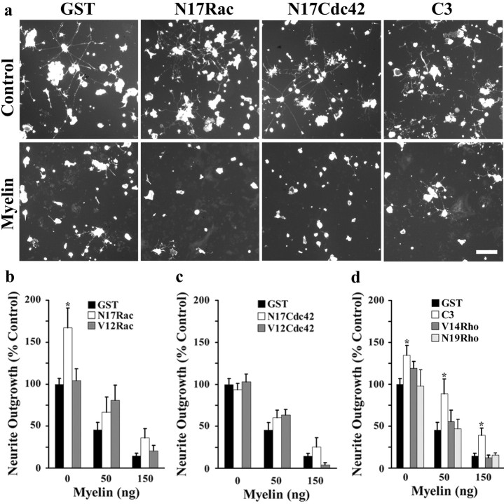
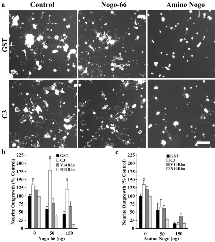
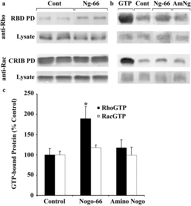
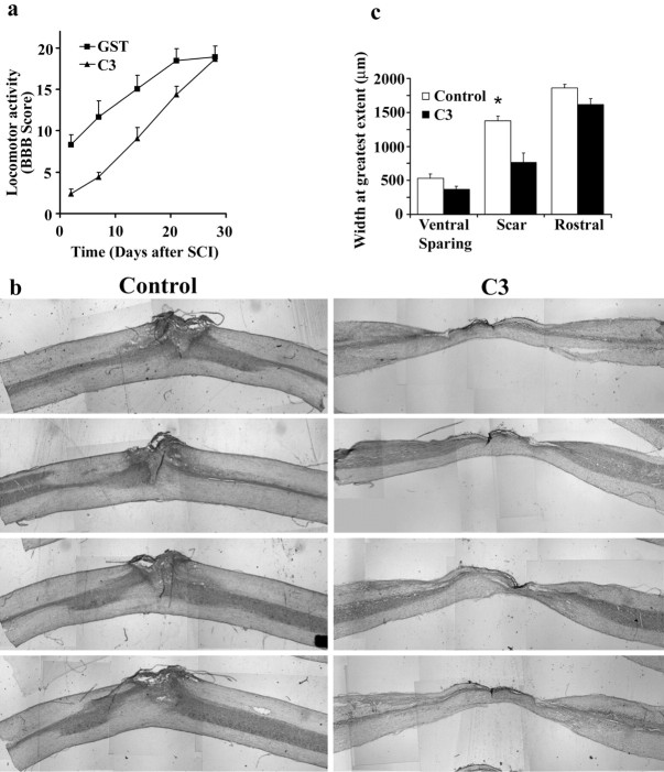
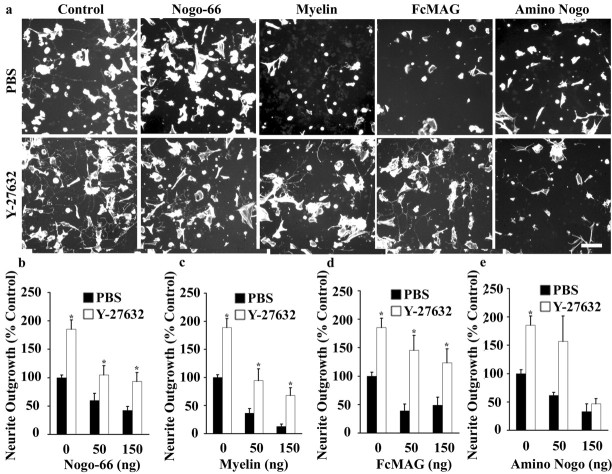
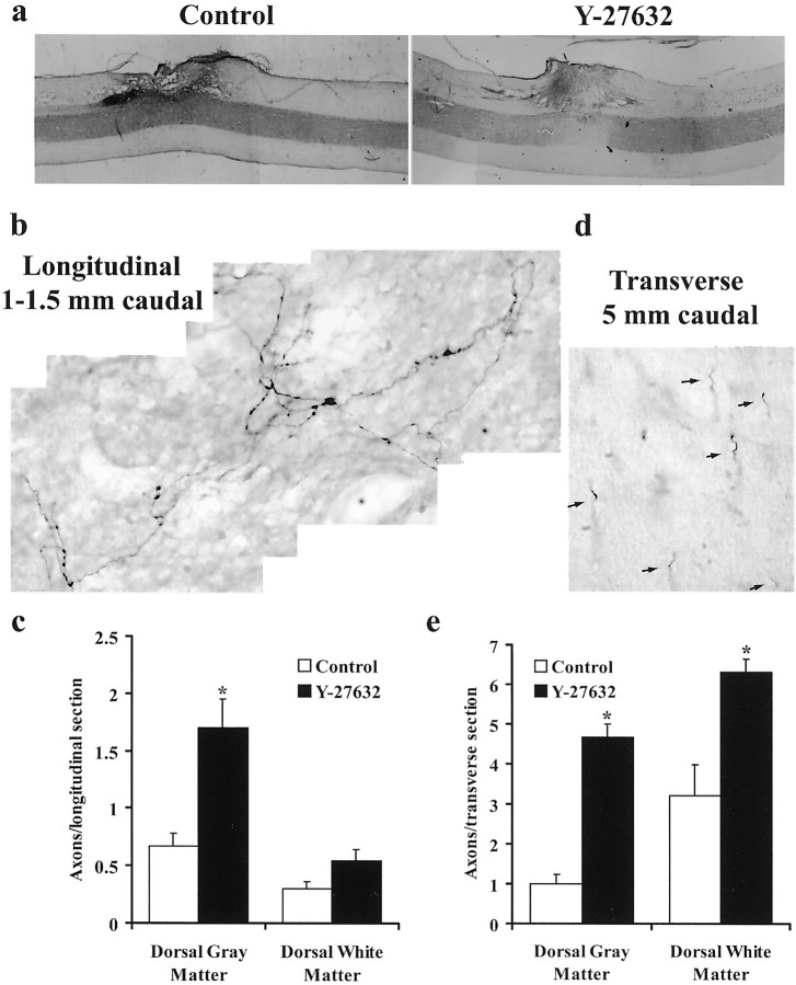
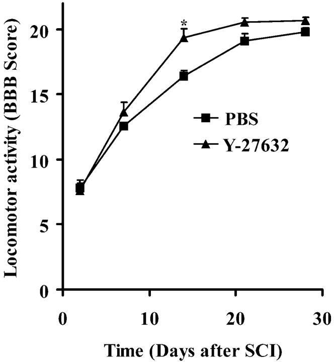
Myelin-associated inhibitors limit axonal regeneration in the injured brain and spinal cord. A common target of many neurite outgrowth inhibitors is the Rho family of small GTPases. Activation of Rho and a downstream effector of Rho, p160ROCK, inhibits neurite outgrowth. Here, we demonstrate that Rho is directly activated by the myelin-associated inhibitor Nogo-66. Using a binding assay to measure Rho activity, we detected increased levels of GTP Rho in PC12 and dorsal root ganglion (DRG) cell lysates after Nogo-66 stimulation. Rho activity levels were not affected by Amino-Nogo stimulation. Rho inactivation with C3 transferase promotes neurite outgrowth of chick DRG neurons in vitro, but with the delivery method used here, it fails to promote neurite outgrowth after corticospinal tract (CST) lesions in the adult rat. Inhibition of p160ROCK with Y-27632 also promotes neurite outgrowth on myelin-associated inhibitors in vitro. Furthermore, Y-27632 enhances sprouting of CST fibers in vivo and accelerates locomotor recovery after CST lesions in adult rats.


Figures

Fig. 1.
Fig. 1.
C3 promotes neurite outgrowth on myelin substrates. a, Dissociated E13 chick DRG neurons were triturated with dominant-negative or constitutively active Rho GTPases or C3 and plated on control or myelin spots. b–d, Quantification of DRG neurite outgrowth per cell on myelin substrates after trituration with Rho GTPases. Determinations are from 3 to 10 separate experiments. Outgrowth is expressed as a percentage of control ± SEM. Scale bar, 100 μm. *p < 0.01 compared with GST treatment.
Fig. 2.
Fig. 2.
C3 promotes neurite outgrowth on Nogo-66.a, Dissociated E13 chick DRG neurons were triturated with GST control protein, Rho, or C3 transferase and plated on control, GST Nogo-66, or Amino–Nogo spots. C3 treatment promotes outgrowth on GST Nogo-66 substrates. Outgrowth remains inhibited on high doses of Amino–Nogo. b, c, Quantification of outgrowth from DRG neurons triturated with GST, C3, or Rho proteins. Determinations are from three to six separate experiments. Outgrowth is expressed as a percentage of control ± SEM. Scale bar, 100 μm. *p < 0.01 compared with GST treatment.
Fig. 3.
Fig. 3.
Nogo-66 directly activates Rho. a, PC12 cells were treated for 30 min with 100 nm GST Nogo-66 (Ng-66), and cell lysates were affinity-precipitated with GST-RBD [RBD pull down(PD)] to detect GTP-bound Rho. GTP Rho levels are enhanced after stimulation with GST Nogo-66. Total Rho protein levels remain unchanged in cell lysates. Lysates were also affinity-precipitated with GST-CRIB (Cdc42/Rac1 interacting and binding domain of p65pak to detect activated Rac. Levels of GTP Rac are unaffected by Nogo-66 stimulation.Cont, Control; b, E13 chick DRGs were plated on control, Nogo-66 (Ng-66), or Amino–Nogo (AmNg) substrates for 3–5 hr. Cell lysates were collected and analyzed for levels of GTP Rho or GTP Rac. Cells plated on Nogo-66 substrates have elevated levels of GTP Rho. As a positive control, cell lysates were incubated for 30 min with GTPγs before the pull-down assay was performed (GTP).c, Quantitation of GTP Rho levels in sensory neurons. GTP Rho levels were normalized to total Rho protein levels in the cell lysate and expressed as a percentage of GTP Rho in control lysates. Determinations are expressed as a percentage of control ± SEM and are from three experiments, each in duplicate. *p = 0.033 compared with the control GTP Rho pull-down assay.
Fig. 4.
Fig. 4.
C3 treatment delays locomotor recovery and disrupts scar formation after rat CST lesions. a, Locomotion in C3-treated rats is delayed compared with GST-treated controls. BBB locomotor assessments were made 2, 7, 14, 21, and 28 d postoperatively. SCI, Spinal cord injury.b, Longitudinal sections of control and C3-treated spinal cords at the site of transection. Note the obvious spinal cord constriction at the lesion site after C3 treatment relative to control.c, Quantification of scar tissue, ventrally spared tissue at the lesion site, and the width of the spinal cord rostral to the lesion. The amount of scar tissue in the C3-treated animals is significantly reduced. Determinations are from nine control animals and nine C3 animals ± SEM. *p < 0.01 compared with vehicle-treated animals.
Fig. 5.
Fig. 5.
Y-27632 promotes neurite outgrowth on inhibitory substrates. a, Dissociated E13 chick DRG neurons were treated with PBS or 10 μm Y-27632 and plated on control, GST Nogo-66, myelin, Fc-MAG, or Amino–Nogo spots. Neurite outgrowth is enhanced on control and inhibitory substrates. Y-27632 does not attenuate the inhibition on high doses of Amino–Nogo.b–e, Quantification of DRG neurite outgrowth on inhibitory substrates in the presence of PBS or Y-27632. Determinations are from 3 to 10 separate experiments. Outgrowth is expressed as a percentage of control ± SEM. Scale bar, 100 μm. *p < 0.01 compared with PBS treatment.
Fig. 6.
Fig. 6.
Y-27632 enhances sprouting of rat CST fibers after dorsal hemisections. a, Longitudinal sections of control and Y-27632-treated spinal cords at the site of transection. The amount of scar tissue, ventral sparing, and spinal cord width rostral to the lesion is similar for Y-27632-treated and control spinal cords.b, Peroxidase staining of BDA-labeled fibers in a longitudinal section 1–1.5 mm caudal to the CST lesion.c, Quantification of the number of axons per longitudinal section caudal to the CST lesion. d, Peroxidase staining of multiple BDA-labeled fibers (arrows) in a transverse section 5 mm caudal to the CST lesion. e, Quantification of the number of axons per transverse section caudal to the CST lesion. Data are from nine vehicle-treated animals and eight Y-27632-treated animals. *p < 0.01 compared with vehicle-treated animals.
Fig. 7.
Fig. 7.
Y-27632 improves locomotor behavior after CST lesions. Rats were evaluated using the BBB score 2, 7, 14, 21, and 28 d postoperatively. *p = 0.01. Data are from 15 vehicle-treated animals and 12 Y-27632-treated animals.SCI, Spinal cord injury.

References

    1. Amano M, Mukai H, Ono Y, Chihara K, Matsui T, Hamajima Y, Okawa K, Iwamatsu A, Kaibuchi K. Identification of a putative target for Rho as the serine-threonine kinase protein kinase N. Science. 1996;271:648–650. - PubMed
    1. Amano M, Chihara K, Kimura K, Fukata Y, Nakamura N, Matsuura Y, Kaibuchi K. Formation of actin stress fibers and focal adhesions enhanced by Rho-kinase. Science. 1997;275:1308–1311. - PubMed
    1. Aspenstrom P, Lindberg U, Hall A. Two GTPases, Cdc42 and Rac, bind directly to a protein implicated in the immunodeficiency disorder Wiskott–Aldrich syndrome. Curr Biol. 1996;6:70–75. - PubMed
    1. Bagrodia S, Taylor SJ, Creasy CL, Chernoff J, Cerione RA. Identification of a mouse p21Cdc42/Rac activated kinase. J Biol Chem. 1995;270:22731–22737. - PubMed
    1. Basso DM, Beattie MS, Bresnahan JC. A sensitive and reliable locomotor rating scale for open field testing in rats. J Neurotrauma. 1995;12:1–21. - PubMed

Publication types

MeSH terms