. 2003 Feb 15;23(4):1424-31.
doi: 10.1523/JNEUROSCI.23-04-01424.2003.

Neurotrophin-3 expressed in situ induces axonal plasticity in the adult injured spinal cord

Affiliations

Neurotrophin-3 expressed in situ induces axonal plasticity in the adult injured spinal cord

Lijun Zhou et al. J Neurosci. .

Abstract

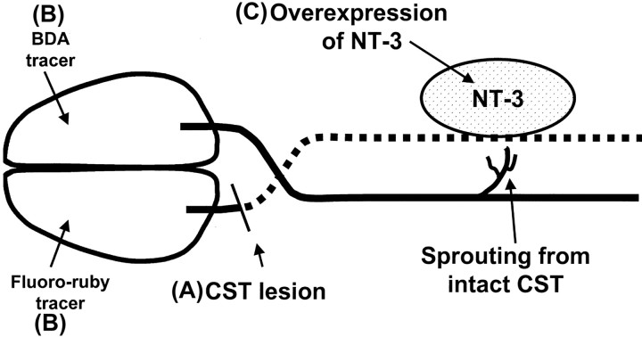
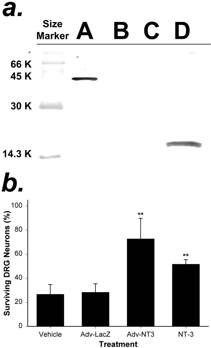
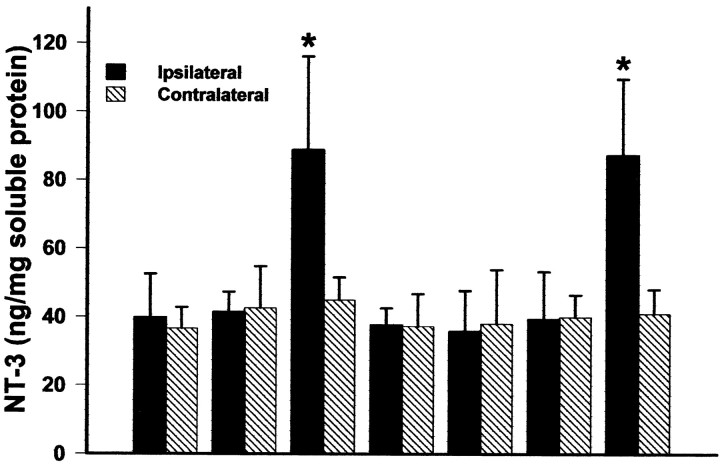
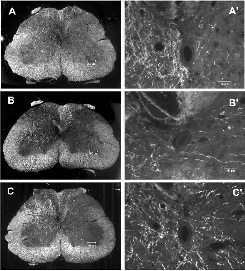
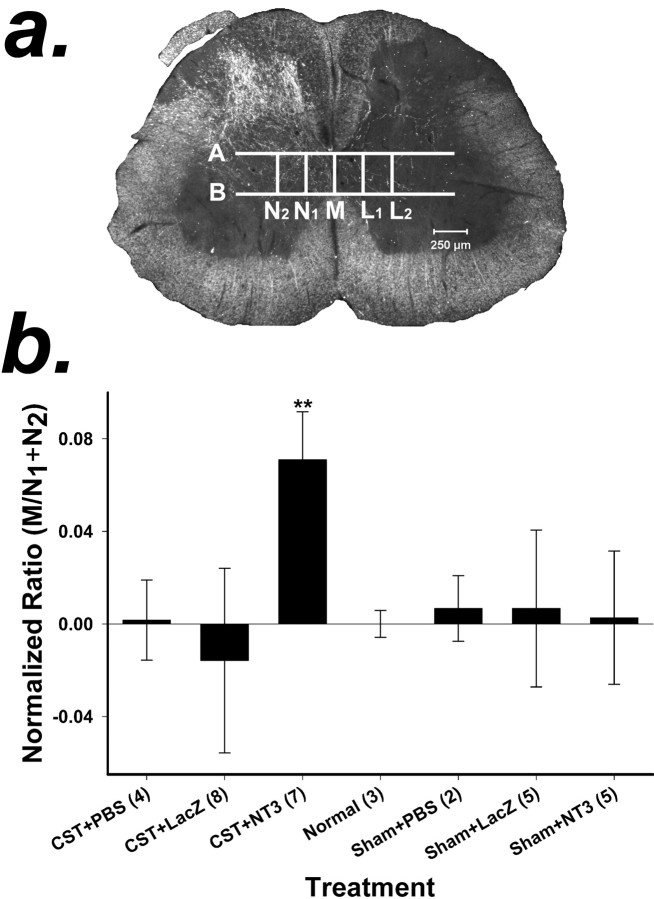
The mammalian CNS lacks the ability to effectively compensate for injury by the regeneration of damaged axons or axonal plasticity of intact axons. However, reports suggest that molecular or cellular manipulations can induce compensatory processes that could support regeneration or plasticity after trauma. We tested whether local, sustained release of the neurotrophic factor neurotrophin-3 (NT-3) would support axonal plasticity in the spinal cord distal to the site of injury in rats. The corticospinal tract (CST) was cut unilaterally at the level of the medulla. This avoided excessive inflammation, secondary cell death, vascular disruption, and the release of inhibitory molecules in the lumbar spinal cord. A replication-defective adenoviral vector (Adv) carrying the NT-3 gene (Adv.EFalpha-NT3) was delivered to the spinal motoneurons by retrograde transport through the sciatic nerve. Retrograde transport of the adenoviral vectors avoided the inflammatory response that would be associated with direct injection into the spinal cord. Transduction of spinal motoneurons with Adv.EFalpha-NT3 resulted in a significant increase in the concentration of NT-3 in the L3-L6 region of the spinal cord for up to 3 weeks. In animals with a CST lesion, this local expression of NT-3 induced growth of axons from the intact CST across the midline to the denervated side. If the CST remained intact, overexpression of NT-3 did not lead to an increase in the number of axons crossing the midline. These data demonstrate that local, sustained expression of NT-3 will support axonal plasticity of intact CST axons after trauma-induced denervation.


Figures

Fig. 1.
Fig. 1.
Schematic of the experimental protocol.A, One CST was lesioned at the level of the hindbrain.B, After 10 d, the neuronal tracers BDA and Fluoro-ruby were injected into opposite sensorimotor cortices.C, Four days after the tracer injection, Adv.EFα-NT3 or Adv.EFα-LacZ was delivered by retrograde transport to the spinal motoneurons in which NT-3 or LacZ were overexpressed.
Fig. 2.
Fig. 2.
Analysis of NT-3 protein produced by cells transduced with Adv.EFα-NT3 in vitro.a, Western blot analysis of the conditioned medium of HeLa cells transduced with Adv. HeLa cells were transduced with Adv.EFα-NT3 or Adv.EFα-LacZ at an MOI of 100. After 12 hr, the medium was replaced with serum-free medium. After 48 hr, the conditioned medium was collected. Western blot analysis using an anti-FLAG antibody was performed to detect NT-3-FLAG in the medium.Lane A, BAP-FLAG; lane B, medium from untransduced HeLa cells; lane C, medium from HeLa cells transduced with Adv.EFα-LacZ; lane D, medium from HeLa cells transduced with Adv.EFα-NT3. A prominent band that cross-reacted with the anti-FLAG antibody is visible inlane D corresponding to the predicted size of the NT-3-FLAG hybrid protein. b, Conditioned medium of HeLa cells transduced with Adv.EFα-NT3 supported the survival of DRG neurons. Primary cultures of DRG neurons were cultured in test media for 48 hr, and the number of surviving neurons was counted in 10 fields. The test media were as follows: Vehicle, 25% conditioned medium from cultures of untransduced HeLa cells;Adv-LacZ, 25% conditioned medium from cultures of Adv.EFα-LacZ-transduced HeLa cells;Adv-NT3, 25% conditioned medium from cultures of Adv.EFα-NT3-transduced HeLa cells; and NT-3, 100 ng of NT-3 protein per milliliter of culture medium. The values are means ± SD of three wells; **p < 0.01 (ANOVA followed by the Student–Newman–Keuls test).
Fig. 3.
Fig. 3.
β-gal is expressed in motoneurons of the lumbar spinal cord. The sciatic nerve was cut, and the proximal stump was inserted into a small chamber filled with Adv.EFα-LacZ(1 × 10⁹ infectious units). After 7 d, rats were perfusion-fixed with 4% PFA, cross sections were cut, and β-gal was identified by histochemical staining with X-gal. β-gal-positive neuronal processes are visible extending from transduced motoneurons (arrowheads) to regions close to the midline and central canal.
Fig. 4.
Fig. 4.
Expression of NT-3 in the L3–L6 lumbar spinal cord 3 weeks after the retrograde delivery of Adv. The concentration of NT-3 in the spinal cord was determined by ELISA (Promega). Values are means ± SD; *p < 0.05;n = 4 per group (ANOVA followed by the Student–Newman–Keuls test).
Fig. 5.
Fig. 5.
A, Photomicrograph at the level of the lesion site in the pyramids showing the extent of the lesioned CST. The nonlesioned CST is demarcated by the anterograde marker BDA stained with ABC reagent (Vector Laboratories) and DAB.B–E, Completeness of the CST lesion demonstrated by anterograde markers in the lumbar spinal cord of unlesioned (B, C) and lesioned (D, E) animals. The unlesioned CST was traced with BDA and visualized with Alexa Fluor 488-conjugated streptavidin; the lesioned CST was traced with Fluoro-ruby.B, Section from a normal rat (sham surgery) showing the unlesioned CST labeled with BDA. C, The same section asB, showing the CST positive for Fluoro-ruby.D, Section from an animal with a complete CSTL, showing the unlesioned side of CST positive for BDA. E, Same section as D showing absence of Fluoro-ruby in the lesioned CST, indicating that the CSTL was complete.
Fig. 6.
Fig. 6.
Sprouting of CST axons across the midline in the spinal cord after CSTL and Adv.EFα-NT3 transduction of motoneurons. Rats with unilateral CSTL were treated with Adv.EFα-NT3 or Adv.EFα-LacZ, whereas the unlesioned CST was labeled with BDA. Dark-field photomicrographs of spinal cord cross sections showed the unlesioned CST axons. A, Section from a normal rat (sham surgery). B, Section from an Adv.EFα-LacZ-treated rat. C, Section from an Adv.EFα-NT3-treated rat. A′–C′, Higher-power photomicrographs of the regions around the central canal.C, BDA-labeled CST neurites can be seen arising from the intact CST, traversing the midline, and growing into the gray matter of the lesioned side of the spinal cord.
Fig. 7.
Fig. 7.
a, Method for quantification of axonal sprouting from the CST across the midline in the spinal cord. Dark-field photomicrographs were taken of each spinal cord section. Five vertical and two horizontal lines were drawn on each photomicrograph of a spinal cord section as reference points for counting axons. M was drawn through the midline,N1 was drawn just lateral to the funiculus of the CST and parallel to M, andN2 was drawn parallel to Mand two times the distance between N1 andM. L1 andL2 were drawn on the lesioned side of the spinal cord and corresponded to lines N1 andN2. Lines A andB were drawn perpendicular to the dorsoventral axis of the cord. A was drawn just below the dorsal columns, andB was drawn just above the ventral columns. Axons that crossed M, N1,N2,L1, and L2within the boundaries of A and B were counted. b, Quantification of CST axons crossing the midline in response to the local expression of NT-3. BDA-positive axons were counted at the midline (M) and at two sites (N1 andN2) in the lateral gray matter of the spinal cord on the side of the unlesioned CST (see Materials and Methods). The ratio of the axons that crossed the midline (M) to those that crossed the two sites in the lateral gray matter (N1 +N2) was computed (M/N1 +N2) to compensate for any variation in the degree of anterograde labeling of the CST. The ratios of the treatment groups were normalized to the ratio of the normal animal group, which was set to zero. A positive value indicates that more axons crossed the midline compared with normal, unlesioned animals; a negative value indicates that fewer axons crossed the midline compared with normal animals. Values are means ± SD; **p < 0.01 (ANOVA followed by the Student–Newman–Keuls test). Numbers inparentheses represent the number of animals per group.

Similar articles

Cited by

References

    1. Baumgartner BJ, Shine HD. Targeted transduction of CNS neurons with adenoviral vectors carrying neurotrophic genes confers neuroprotection that exceeds the transduced population. J Neurosci. 1997;17:6504–6511. - PMC - PubMed
    1. Baumgartner BJ, Shine HD. Neuroprotection of spinal motoneurons following targeted transduction with an adenoviral vector carrying the gene for glial cell-line derived neurotrophic factor. Exp Neurol. 1998a;153:102–112. - PubMed
    1. Baumgartner BJ, Shine HD. Permanent rescue of lesioned neonatal motoneurons and enhanced axonal regeneration by adenovirus-mediated expression of glial cell-line-derived neurotrophic factor. J Neurosci Res. 1998b;54:766–777. - PubMed
    1. Benowitz LI, Goldberg DE, Madsen JR, Soni D, Irwin N. Inosine stimulates extensive axon collateral growth in the rat corticospinal tract after injury. Proc Natl Acad Sci USA. 1999;96:13486–13490. - PMC - PubMed
    1. Brösamle C, Schwab ME. Cells of origin, course, and termination patterns of the ventral, uncrossed component of the mature rat corticospinal tract. J Comp Neurol. 1997;386:293–303. - PubMed

Publication types

MeSH terms