. 2003 Feb 15;23(4):1478-86.
doi: 10.1523/JNEUROSCI.23-04-01478.2003.

A novel functional neuron group for respiratory rhythm generation in the ventral medulla

Affiliations

A novel functional neuron group for respiratory rhythm generation in the ventral medulla

Hiroshi Onimaru et al. J Neurosci. .

Abstract

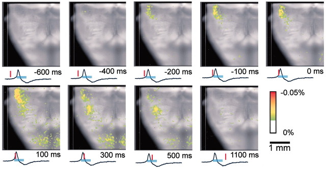
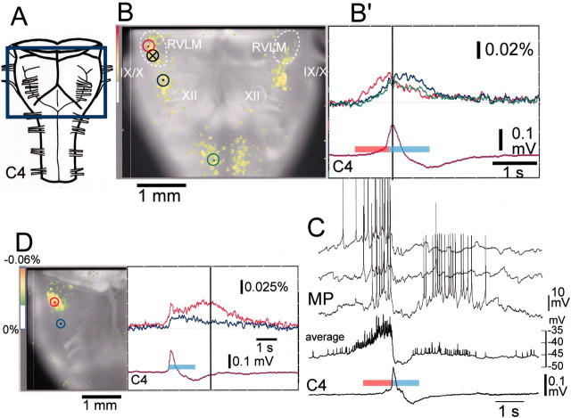
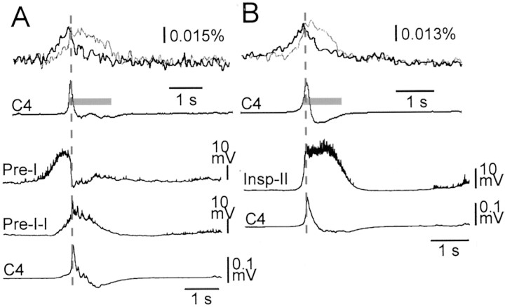
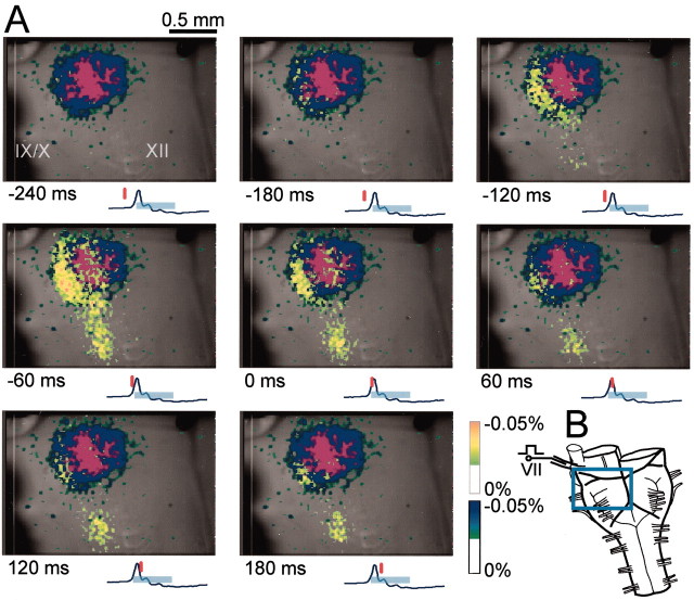
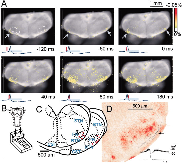
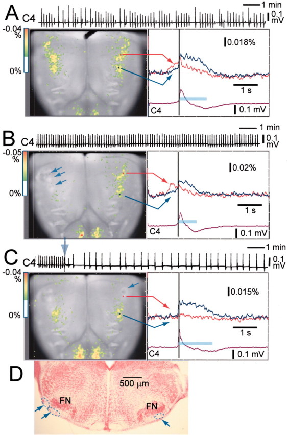
We visualized respiratory neuron activity covering the entire ventral medulla using optical recordings in a newborn rat brainstem-spinal cord preparation stained with voltage-sensitive dye. We measured optical signals from several seconds before to several seconds after the inspiratory phase using the inspiratory motor nerve discharge as the trigger signal; we averaged the optical signals of 50-150 respiratory cycles to obtain an optical image correlating particularly to inspiratory activity. The optical images we obtained from the ventral approach indicated that neuron activity first appeared during the respiratory cycle in the limited region of the rostral ventrolateral medulla (RVLM), preceding the onset of inspiratory activity by approximately 500 msec. During the inspiratory phase, plateau activity appeared in the more caudal ventrolateral medulla at the level of the most rostral roots of the XIIth nerve. Comparison with electrophysiological recordings from respiratory neurons in the RVLM suggested that the optical signals preceding the inspiratory burst reflect preinspiratory neuron activity in this area. This RVLM area was determined to be ventrolateral to the facial nucleus and close to the ventral surface. We referred to this functional neuron group as the para-facial respiratory group (pFRG). Partial, bilateral electrical lesioning of the pFRG significantly reduced the respiratory frequency, together with changes in the spatiotemporal pattern of respiratory neuron activity. Our findings suggest that the pFRG comprises a neuronal population that is involved in the primary respiratory rhythm generation in the rostrocaudally extending respiratory neuron network of the medulla.


Figures

Fig. 1.
Fig. 1.
Spatiotemporal pattern of respiratory neuron activity observed on optical images. The image is superimposed on the ventral surface of the right half of the medulla. Also see Figure 2 for the orientation of the preparation. The numbers at thebottom right of each image denote the time from the start of C4 inspiratory activity. The tracing under each image is the C4 activity; the inspiratory phase is indicated by the light-blue bar. The red vertical lineon the C4 tracing shows the time at which the image was obtained. Results are the average of 100 respiratory cycles triggered by C4 inspiratory activity. The preparation was stained with 50 μg/ml Di-2-ANEPEQ for 40 min. The sampling clock is 20 msec. Note that optical signals reflecting respiratory neuron activity appear in the RVLM during the preinspiratory phase. During the inspiratory phase, peak activity appears in the more caudal ventrolateral medulla.
Fig. 2.
Fig. 2.
Fluorescence changes in the optical recording and electrophysiological recordings of respiratory neuron activity. The preparation is the same as in Figure 1 except for D.A, Ventral aspect of the preparation and the optical image recording area boxed in blue.B, Optical image of respiratory neuron activity near the C4 peak (at dark vertical line in B′) superimposed on the ventral surface of the medulla. IX/X, XII, Cranial nerves; white dotted lines, approximate area of the RVLM; B′, Fluorescence changes at three different points indicated by dots inred, blue, and green circles on B: red, in the RVLM at the level just rostral to the IX/Xth cranial roots;blue, in the more caudal ventrolateral medulla at the level of the rostral roots of the XIIth cranial nerve; andgreen, in the ventral horns of the cervical cord. Fluorescence decrease (i.e., depolarization) is upward.C4, Electrical record of C4 inspiratory activity. Thered bar and light-blue bar on the C4 tracing denote the preinspiratory and inspiratory phases, respectively. Note that the fluorescence change precedes the inspiratory phase in all of the areas indicated, whereas the amplitude is larger in the RVLM (red tracing). C, Membrane potential trajectory (MP) of a Pre-I neuron recorded in the ventrolateral medulla 1.4 mm lateral to the midline and at the level of the IX/Xth roots of the same preparation (circle with cross in B). Three membrane potential traces are shown, one below the other. The averages of the membrane potential trajectory and C4 activity for 21 respiratory cycles are also shown (average, C4). The red bar and light-blue bar on the average C4 tracing denote the preinspiratory and inspiratory phases, respectively.D, An example exhibiting clear postinspiratory activity.Left, Optical image of respiratory neuron activity in the postinspiratory phase (dark vertical line atright) superimposed on the ventral surface of the right-side medulla. Right, Fluorescence changes at two different points indicated by dots inred and blue circles on the left image: red, in the RVLM at the level just rostral to the IX/Xth cranial roots; and blue, in the more caudal ventrolateral medulla near the level of the rostral roots of the XIIth cranial nerve. Results are the average of 100 respiratory cycles triggered by C4 inspiratory activity. The preparation was stained with 0.2 mg/ml Di-4-ANEPPS for 30 min. The sampling clock is 25 msec. Note the short (75 msec) preinspiratory activity and long (∼2 sec) postinspiratory activity.
Fig. 3.
Fig. 3.
Comparison between fluorescence changes in optical recordings and averaged membrane potential trajectories of different types of respiratory neurons. A, B, top, Fluorescence changes at two different points similar to Figure2B or D: thick line, in the RVLM at the level just rostral to the IX/Xth cranial roots; thin line, in the more caudal ventrolateral medulla near the level of the rostral roots of the XIIth cranial nerve. Results are the average of 50 (in A) or 100 (in B) respiratory cycles triggered by C4 inspiratory activity. Fluorescence decrease (i.e., depolarization) is upward. The preparation was stained with 50 μg/ml Di-2-ANEPEQ for 45 min in A or 43 min in B. The sampling clock is 20 msec. Gray bars on the average C4 tracing (C4) denote the inspiratory phase. A, B, bottom, Averaged membrane potential trajectories from a Pre-I neuron, a Pre-I–I type inspiratory neuron, and a type II inspiratory neuron (Insp-II), recorded in the ventrolateral medulla 1.3–1.4 mm lateral to the midline and at the level of the IX/Xth roots. C4, Averaged C4 activity. Thevertical dotted lines in A andB denote the time of peak C4 activity.
Fig. 4.
Fig. 4.
Optical images of respiratory neuron activity superimposed on the area of the facial nucleus.A, The dark blue area in each image indicates the facial nucleus of the right side of the medulla, which was activated antidromically by stimulation of the right facial nerve.Purple denotes a fluorescence change below -0.05%. Thenumbers at the bottom left of each image denote the time from the start of C4 inspiratory activity. Thetracing under each image is the C4 activity; the inspiratory phase is indicated by the light-blue bar. The red vertical line on the C4 tracing shows the time at which the image was obtained. The preparation was stained with 50 μg/ml Di-2-ANEPEQ for 40 min. Results are the average of 50 respiratory cycles triggered by C4 inspiratory activity. The sampling clock is 30 msec. Note that optical signals during the preinspiratory phase appear to overlap to the lateral edge of the facial nucleus.IX/X, XII, Cranial nerves. B, Diagram of the ventral aspect of the preparation and the optical image recording area boxed in blue. VII, Facial nerve.
Fig. 5.
Fig. 5.
Optical images of respiratory neuron activity obtained by observation from the cut surface of a cross section of the rostral medulla. A, Spatiotemporal pattern of optical images of respiratory neuron activity superimposed on the cut surface of the rostral medulla. The numbers at the bottom right of each image denote the time from the start of C4 inspiratory activity. The tracing under each image is the C4 activity; the inspiratory phase is indicated by thelight-blue bar. The vertical red line on the C4 tracing shows the time at which the top image was obtained. Results are the average of 100 respiratory cycles triggered by C4 inspiratory activity. The sampling clock is 20 msec. Note that optical signals reflecting respiratory neuron activity appear in the part ventrolateral to the facial nucleus (top left, white dotted line) during the preinspiratory phase (arrows). B, Schematic representation of the preparation. The preparation was cut transversely at the level (indicated by a dotted line) rostral to the IX/Xth cranial nerve roots and then stained with 0.1 mg/ml Di-4-ANEPPS for 56 min. It was then fixed onto a rubber block with pins and cut surface up, for observation from the cut surface of the cross section (arrows). C, Pre-I neurons in the part ventrolateral to the facial nucleus. The neuron activities were recorded in the whole-cell configuration in the same type of preparations as illustrated in B by approaching from the rostral cut surface. Red dots indicate locations of recorded neurons. Neurons were found in the reticular formation ventrolateral to the facial nucleus, and some neurons were also found in the ventrolateral edge of the facial nucleus.Dotted lines indicate demarcation between some anatomical structures. FN, Facial nucleus;CST, corticospinal tract; STN, spinal trigeminal nucleus, oral part. D, An example of reconstruction of a Pre-I neuron stained with LY. Thearrow denotes the site of soma located ventrolateral to the facial nucleus. The axon could not be traced. The averaged membrane potential trajectory is shown in the bottom right with C4 activity.
Fig. 6.
Fig. 6.
Effects of partial electrical lesioning of the RVLM on C4 rhythm and spatiotemporal pattern of respiratory neuron activity. A, C4 activity and optical records in control.B, C4 activity and optical records after partial lesioning of three points (arrows) in the right side of the RVLM. C, C4 activity and optical records after subsequent partial lesioning of a point (arrow) in the left side of a similar rostral medulla. C4, Inspiratory C4 activity. A–C, left, Optical images of respiratory neuron activity near the C4 onset (dark vertical line atright) superimposed on the ventral surface of the medulla. A–C, right, Fluorescence changes at two points indicated on each right panel: red, in the RVLM at the level just rostral to the IX/Xth cranial roots;blue, in the more caudal ventrolateral medulla at the level of the rostral roots of the XIIth cranial nerve; the inspiratory phase is indicated by the light-blue bar. Fluorescence decrease (i.e., depolarization) is upward. The preparation was stained with 50 μg/ml Di-2-ANEPEQ for 50 min. Results are the average of 50 respiratory cycles triggered by C4 inspiratory activity. The sampling clock is 20 msec. Note that preinspiratory activity recorded optically in the intact side is still clearly observed after partial lesioning of the right side of the rostral medulla (B). After subsequent partial lesioning of the other side, preinspiratory activity recorded optically is markedly depressed and the C4 rate is reduced (arrow on the C4 record), whereas the duration of inspiratory activity in the caudal medulla as well as that of C4 activity are prolonged. D, Histological verification of the electrical lesioning; 100 μm transverse section. The lesions indicated by dotted lines andarrows are confirmed in the limited area ventrolateral to the facial nucleus (FN).

Similar articles

Cited by

References

    1. Arata A, Onimaru H, Homma I. Respiration-related neurons in the ventral medulla of newborn rats in vitro. Brain Res Bull. 1990;24:599–604. - PubMed
    1. Ballanyi K, Onimaru H, Homma I. Respiratory network function in the isolated brainstem–spinal cord of newborn rats. Prog Neurobiol. 1999;59:583–634. - PubMed
    1. Bianchi AL, Denavit-Saubie M, Champagnat J. Central control of breathing in mammals: neuronal circuitry, membrane properties, and neurotransmitters. Physiol Rev. 1995;75:1–45. - PubMed
    1. Bodineau L, Cayetanot F, Frugiere A. Possible role of retrotrapezoid nucleus and parapyramidal area in the respiratory response to anoxia: an in vitro study in neonatal rat. Neurosci Lett. 2000;295:67–69. - PubMed
    1. Cohen LB, Lesher S. Optical monitoring of membrane potential: methods of multisite optical measurement. In: De Weer P, Salzberg BM, editors. Optical methods in cell physiology. Wiley; New York: 1986. pp. 71–99. - PubMed

Publication types

Substances

LinkOut - more resources