

Changes in inhibitory amino acid release linked to pontine-induced atonia: an in vivo microdialysis study
- PMID: 12598643
- PMCID: PMC6742274
- DOI: 10.1523/JNEUROSCI.23-04-01548.2003
Changes in inhibitory amino acid release linked to pontine-induced atonia: an in vivo microdialysis study
Abstract
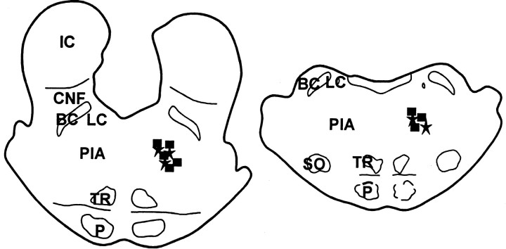
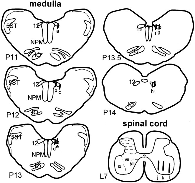
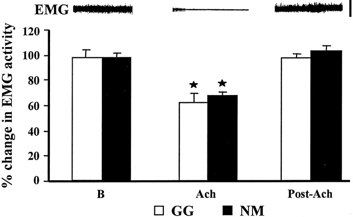
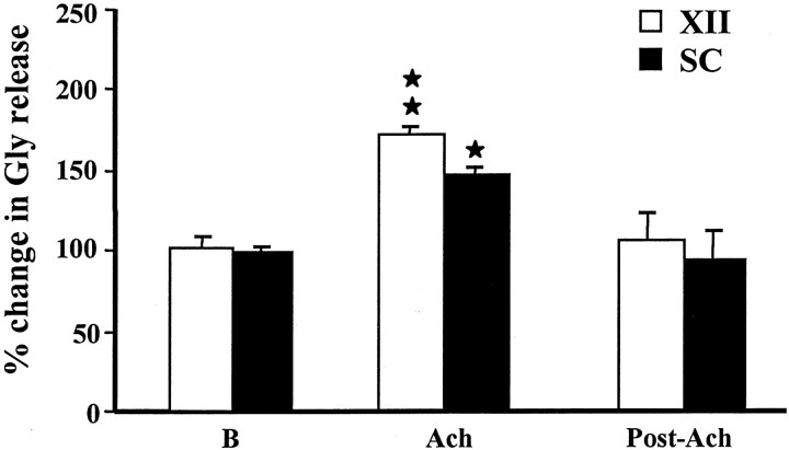
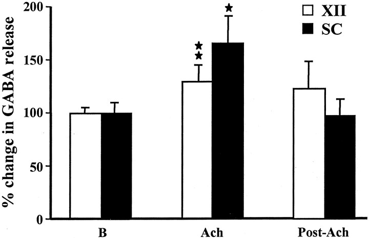
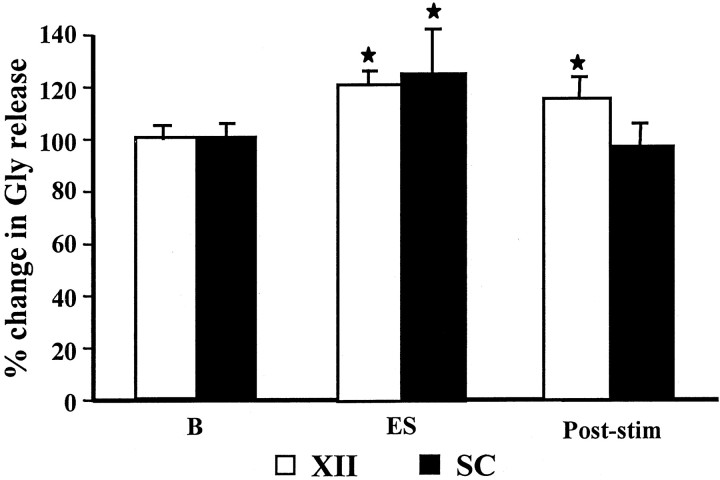
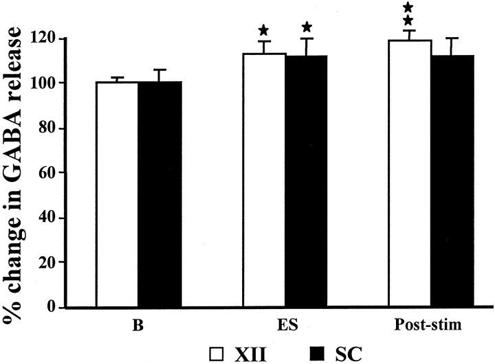
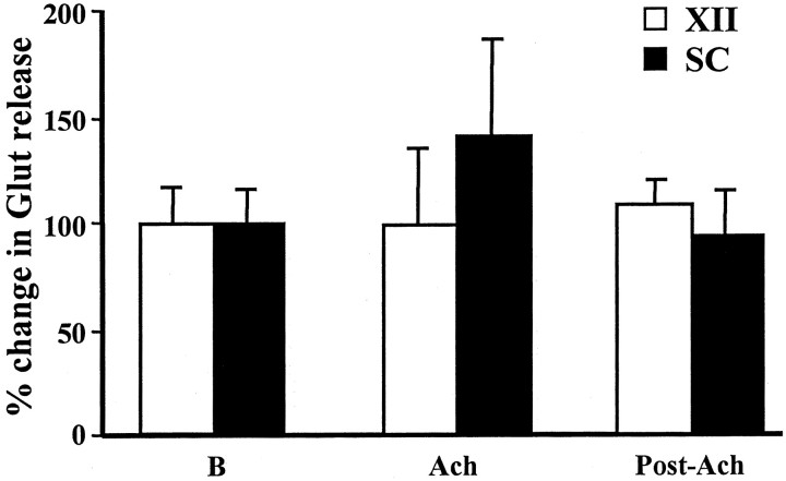
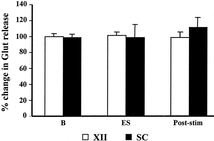
We hypothesized that cessation of brainstem monoaminergic systems and an activation of brainstem inhibitory systems are both involved in pontine inhibitory area (PIA) stimulation-induced muscle atonia. In our previous study (Lai et al., 2001), we found a decrease in norepinephrine and serotonin release in motoneuron pools during PIA stimulation-induced muscle tone suppression. We now demonstrate an increase in inhibitory amino acid release in motor nuclei during PIA stimulation in the decerebrate cat using in vivo microdialysis and HPLC analysis techniques. Microinjection of acetylcholine into the PIA elicited muscle atonia and simultaneously produced a significant increase in both glycine and GABA release in both the hypoglossal nucleus and the lumbar ventral horn. Glycine release increased by 74% in the hypoglossal nucleus and 50% in the spinal cord. GABA release increased by 31% in the hypoglossal nucleus and 64% in the spinal cord during atonia induced by cholinergic stimulation of the PIA. As with cholinergic stimulation, 300 msec train electrical stimulation of the PIA elicited a significant increase in glycine release in the hypoglossal nucleus and ventral horn. GABA release was significantly increased in the hypoglossal nucleus but not in the spinal cord during electrical stimulation of the PIA. Glutamate release in the motor nuclei was not significantly altered during atonia induced by electrical or acetylcholine stimulation of the PIA. We suggest that both glycine and GABA play important roles in the regulation of upper airway and postural muscle tone. A combination of decreased monoamine and increased inhibitory amino acid release in motoneuron pools causes PIA-induced atonia and may be involved in atonia linked to rapid eye-movement sleep.
Figures
Similar articles
-
Changes in monoamine release in the ventral horn and hypoglossal nucleus linked to pontine inhibition of muscle tone: an in vivo microdialysis study.J Neurosci. 2001 Sep 15;21(18):7384-91. doi: 10.1523/JNEUROSCI.21-18-07384.2001. J Neurosci. 2001. PMID: 11549748 Free PMC article.
-
Changes in serotonin level in the hypoglossal nucleus region during carbachol-induced atonia.Brain Res. 1994 May 9;645(1-2):291-302. doi: 10.1016/0006-8993(94)91663-2. Brain Res. 1994. PMID: 7520343
-
Suppression of hypoglossal motoneurons during the carbachol-induced atonia of REM sleep is not caused by fast synaptic inhibition.Brain Res. 1993 May 21;611(2):300-12. doi: 10.1016/0006-8993(93)90517-q. Brain Res. 1993. PMID: 8334524
-
Contribution of postural muscle tone to full expression of posture and locomotor movements: multi-faceted analyses of its setting brainstem-spinal cord mechanisms in the cat.Jpn J Physiol. 1989;39(6):785-809. Jpn J Physiol. 1989. PMID: 2698966 Review.
-
New aspects in the pathophysiology of rapid eye movement sleep behavior disorder: the potential role of glutamate, gamma-aminobutyric acid, and glycine.Sleep Med. 2013 Aug;14(8):714-8. doi: 10.1016/j.sleep.2013.02.004. Epub 2013 Jun 19. Sleep Med. 2013. PMID: 23790501 Review.
Cited by
-
Illuminating the locus coeruleus: control of posture and arousal.Nat Neurosci. 2010 Dec;13(12):1448-9. doi: 10.1038/nn1210-1448. Nat Neurosci. 2010. PMID: 21102568 Free PMC article.
-
Unearthing the phylogenetic roots of sleep.Curr Biol. 2008 Aug 5;18(15):R670-R679. doi: 10.1016/j.cub.2008.06.033. Curr Biol. 2008. PMID: 18682212 Free PMC article. Review.
-
Neurobiology of waking and sleeping.Handb Clin Neurol. 2011;98:131-49. doi: 10.1016/B978-0-444-52006-7.00009-5. Handb Clin Neurol. 2011. PMID: 21056184 Free PMC article. Review. No abstract available.
-
The anatomical, cellular and synaptic basis of motor atonia during rapid eye movement sleep.J Physiol. 2016 Oct 1;594(19):5391-414. doi: 10.1113/JP271324. Epub 2016 Jul 3. J Physiol. 2016. PMID: 27060683 Free PMC article. Review.
-
Neuroanatomical Basis of State-Dependent Activity of Upper Airway Muscles.Front Neurol. 2018 Sep 10;9:752. doi: 10.3389/fneur.2018.00752. eCollection 2018. Front Neurol. 2018. PMID: 30250449 Free PMC article. Review.
References
-
- Aldes LD, Chronister RB, Marco LA. Distribution of glutamic acid decarboxylase and gamma-aminobutyric acid in the hypoglossal nucleus in the rat. J Neurosci Res. 1988;19:343–348. - PubMed
-
- Berman AL. The brain stem of the cat. University of Wisconsin; Madison, WI: 1968.
-
- Chirwa SS, Stafford-Segert I, Soja PJ, Chase MH. Strychnine antagonizes jaw-closer motoneuron IPSPs induced by reticular stimulation during active sleep. Brain Res. 1991;547:323–326. - PubMed
Publication types
MeSH terms
Substances
Grants and funding
LinkOut - more resources
Full Text Sources
Other Literature Sources
Research Materials
Miscellaneous