

Evidence for rotation of V1-ATPase
- PMID: 12598655
- PMCID: PMC151337
- DOI: 10.1073/pnas.0436796100
Evidence for rotation of V1-ATPase
Abstract
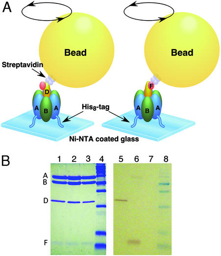
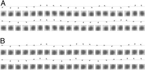
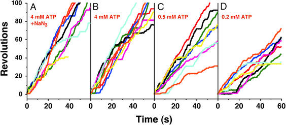
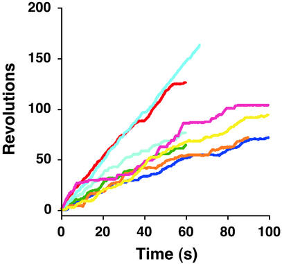
V(o)V(1)-ATPase is responsible for acidification of eukaryotic intracellular compartments and ATP synthesis of Archaea and some eubacteria. From the similarity to F(o)F(1)-ATP synthase, V(o)V(1)-ATPase has been assumed to be a rotary motor, but to date there are no experimental data to support this. Here we visualized the rotation of single molecules of V(1)-ATPase, a catalytic subcomplex of V(o)V(1)-ATPase. V(1)-ATPase from Thermus thermophilus was immobilized onto a glass surface, and a bead was attached to the D or F subunit through the biotin-streptavidin linkage. In both cases we observed ATP-dependent rotations of beads, the direction of which was always counterclockwise viewed from the membrane side. Given that three ATP molecules are hydrolyzed per one revolution, rates of rotation agree consistently with rates of ATP hydrolysis at saturating ATP concentrations. This study provides experimental evidence that V(o)V(1)-ATPase is a rotary motor and that both D and F subunits constitute a rotor shaft.
Figures
References
-
- Nishi T, Forgac M. Nat Rev Mol Cell Biol. 2002;3:94–103. - PubMed
-
- Yoshida M, Muneyuki E, Hisabori T. Nat Rev Mol Cell Biol. 2001;2:669–677. - PubMed
-
- Boyer P D. Biochim Biophys Acta. 1993;1140:215–250. - PubMed
-
- Stevens T H, Forgac M. Annu Rev Cell Dev Biol. 1997;13:779–808. - PubMed
-
- Yokoyama K, Oshima T, Yoshida M. J Biol Chem. 1990;265:21946–21950. - PubMed
Publication types
MeSH terms
Substances
LinkOut - more resources
Full Text Sources
Other Literature Sources
