

Bilateral brain abnormalities associated with dominantly inherited verbal and orofacial dyspraxia
- PMID: 12599277
- PMCID: PMC6872113
- DOI: 10.1002/hbm.10093
Bilateral brain abnormalities associated with dominantly inherited verbal and orofacial dyspraxia
Abstract
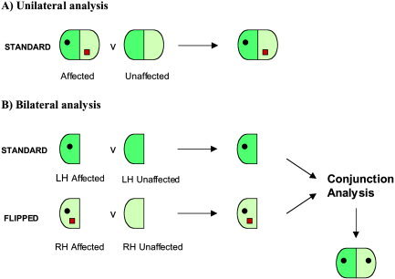
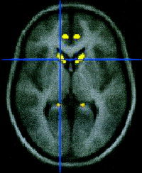
The KE family is a large three-generational pedigree in which half of the members suffer from a verbal and orofacial dyspraxia in association with a point mutation in the FOXP2 gene. This report extends previous voxel-based morphometric analyses of magnetic resonance imaging (MRI) scans (Watkins et al. [2002] Brain 125:465-478) using a bilateral conjunction analysis. This searches specifically for areas of grey matter density that differ bilaterally in the affected members compared with both matched controls and the unaffected family members. 3-D T1-weighted MRI datasets of 17 family members (10 affected, 7 unaffected) and matched controls were compared. The most significant findings were reduced grey matter density bilaterally in the caudate nucleus, the cerebellum, and the left and right inferior frontal gyrus in the affected members. In addition, increased grey matter density was found bilaterally in the planum temporale. These results confirm that a point mutation in FOXP2 is associated with several bilateral grey matter abnormalities in both motor and language related regions. The results also demonstrate the advantages of using a conjunction analysis when bilateral abnormalities are suspected.
The KE family is a large three‐generational pedigree in which half of the members suffer from a verbal and orofacial dyspraxia in association with a point mutation in the FOXP2 gene. This report extends previous voxel‐based morphometric analyses of magnetic resonance imaging (MRI) scans (Watkins et al. [2002] Brain 125:465–478) using a bilateral conjunction analysis. This searches specifically for areas of grey matter density that differ bilaterally in the affected members compared with both matched controls and the unaffected family members. 3‐D T1‐weighted MRI datasets of 17 family members (10 affected, 7 unaffected) and matched controls were compared. The most significant findings were reduced grey matter density bilaterally in the caudate nucleus, the cerebellum, and the left and right inferior frontal gyrus in the affected members. In addition, increased grey matter density was found bilaterally in the planum temporale. These results confirm that a point mutation in FOXP2 is associated with several bilateral grey matter abnormalities in both motor and language related regions. The results also demonstrate the advantages of using a conjunction analysis when bilateral abnormalities are suspected. Hum. Brain Mapping 18:194–200, 2003. © 2003 Wiley‐Liss, Inc.
Copyright 2003 Wiley-Liss, Inc.
Figures
References
-
- Abdullaev YG, Bechtereva NP, Melnichuk KV (1998): Neuronal activity of human caudate nucleus and prefrontal cortex in cognitive tasks. Behav Brain Res 97: 159–177. - PubMed
-
- Ashburner J, Friston KJ (2000): Voxel‐based morphometry: the methods. NeuroImage 11: 805–821. - PubMed
-
- Diamond A (2000): Close interrelation of motor development and cognitive development and of the cerebellum and prefrontal cortex. Child Dev 71: 44–56. - PubMed
-
- Fisher SE, Vargha‐Khadem F, Watkins KE, Monaco AP, Pembrey ME (1998): Localisation of a gene implicated in a severe speech and language disorder. Nat Genet 18: 168–170. - PubMed
Publication types
MeSH terms
Substances
Grants and funding
LinkOut - more resources
Full Text Sources
Other Literature Sources
