. 2003 Feb 24:3:1.
doi: 10.1186/1471-5945-3-1. Epub 2003 Feb 24.

Psoriasin (S100A7) expression is altered during skin tumorigenesis

Affiliations

Psoriasin (S100A7) expression is altered during skin tumorigenesis

Salem Alowami et al. BMC Dermatol. .

Abstract

Background: Psoriasin (S100A7) expression has previously been associated with psoriasiform hyperplasia as well as with tumor progression in breast cancer. Its expression profile for different stages of skin lesions is unknown. The aim of this study was to determine the relationship between psoriasin (S100A7) and tumor progression in skin.

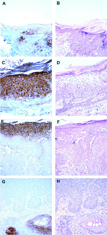
Methods: Psoriasin was assessed by immunohistochemistry and levels of expression determined by semi-quantitative scoring in skin biopsies from 50 patients. The cohort included normal skin, actinic keratosis, squamous carcinoma in-situ, invasive squamous cell carcinoma, and basal cell carcinoma.

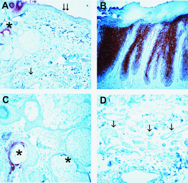
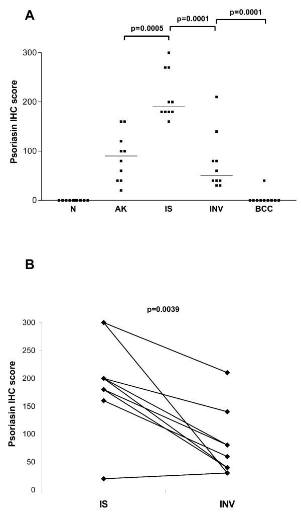
Results: In normal skin, psoriasin was rarely detected in epidermis but was expressed in underlying adnexae. In abnormal epidermis psoriasin was frequently expressed in abnormal keratinocytes in actinic keratosis, in-situ and invasive squamous cell carcinoma, but was rarely observed in the basal epidermal layer or in superficial or invasive basal cell carcinoma. The highest levels of expression were seen within squamous carcinoma in-situ. Significantly reduced levels of expression were observed in both unmatched (p = 0.0001) and matched (p < 0.004) invasive squamous cell carcinoma. Psoriasin expression within abnormal squamous lesions correlated with mitotic count (r = 0.54, p = 0.0036), however no significant relation was found with the intensity of dermal inflammatory cell infiltrates assessed within each pathology.

Conclusion: These results suggest that altered psoriasin expression occurs in abnormal epidermis and that downregulation may be related to the onset of invasion in squamous cell carcinoma in skin.


Figures

Figure 1
Figure 1
Immunohistochemical detection of psoriasin in normal and psoriatic skin Pattern of psoriasin expression in normal skin and psoriatic lesions detected by immunohistochemistry. Psoriasin is absent from normal dermis, epidermis (A, double arrow), and eccrine glands (A & D, single arrow) but is expressed in hair follicles and occasional associated sebaceous glands (A & C, asterix). Psoriasin is highly expressed in superficial layers of epidermis within psoriatic lesions (B). Original magnification A&B 200×, C&D 400×.
Figure 2
Figure 2
Immunohistochemical detection of psoriasin in skin Pattern of psoriasin expression in pre-neoplastic and neoplastic skin lesions detected by immunohistochemistry (panels A,C,E,F) with corresponding areas from H&E stains (panels B,D,F,H). Psoriasin is moderately (A, right) or highly (C) expressed in carcinoma in-situ alone and carcinoma in-situ associated with invasive carcinoma (E, upper), and downregulated in invasive squamous carcinoma (E, lower). Psoriasin is absent in invasive basal cell carcinoma (G, upper) in comparison with adjacent positively staining normal hair follicles (G, lower right). Original magnification, 200×.
Figure 3
Figure 3
Psoriasin expression relative to lesion type Panel A: distribution of expression within cases with normal skin (N), actinic keratosis (AK), squamous carcinoma in-situ (IS), invasive squamous cell carcinoma (INV), and basal cell carcinoma (BCC). Bars represent medians. All p values were determined by Mann Whitney test. Panel B: comparison of expression within individual cases between areas of squamous carcinoma in-situ (IS) and invasive squamous cell carcinoma (INV). The p value was determined by Wilcoxon Rank Sum test.
Figure 4
Figure 4
Psoriasin expression relative to mitosis and inflammation within lesion type. Panel A: Psoriasin expression levels determined by IHC score relative to mitotic count in squamous epithelial pathologies (actinic keratosis, squamous carcinoma in-situ and squamous invasive lesions). Panel B: Psoriasin expression levels determined by IHC score relative to inflammation within each lesion type. Distribution of expression within cases without inflammation (-) and with inflammation (+) for actinic keratosis (AK-, AK+), squamous carcinoma in-situ (IS-, IS+), and invasive squamous cell carcinoma (INV-, INV+). Bars represent medians. P values determined by Mann Whitney test.

References

    1. Heizmann CW, Fritz G, Schafer BW. S100 proteins: structure, functions and pathology. Front Biosci. 2002;7:d1356–d1368. - PubMed
    1. Madsen P, Rasmussen HH, Leffers H, Honore B, Dejgaard K, Olsen E, et al. Molecular cloning, occurrence, and expression of a novel partially secreted protein "psoriasin" that is highly up-regulated in psoriatic skin. J Invest Dermatol. 1991;97:701–712. - PubMed
    1. Algermissen B, Sitzmann J, LeMotte P, Czarnetzki B. Differential expression of CRABP II, psoriasin and cytokeratin 1 mRNA in human skin diseases. Arch Dermatol Res. 1996;288:426–430. doi: 10.1007/s004030050077. - DOI - PubMed
    1. Jinquan T, Vorum H, Larsen CG, Madsen P, Rasmussen HH, Gesser B, et al. Psoriasin: a novel chemotactic protein. J Invest Dermatol. 1996;107:5–10. - PubMed
    1. Van Ruissen F, de Jongh GJ, Zeeuwen PL, Van Erp PE, Madsen P, Schalkwijk J. Induction of normal and psoriatic phenotypes in submerged keratinocyte cultures. J Cell Physiol. 1996;168:442–452. doi: 10.1002/(SICI)1097-4652(199608)168:2<442::AID-JCP23>3.0.CO;2-3. - DOI - PubMed

Publication types