. 2003 Mar;121(3):215-25.
doi: 10.1085/jgp.20028760.

Rapid induction of P/C-type inactivation is the mechanism for acid-induced K+ current inhibition

Affiliations

Rapid induction of P/C-type inactivation is the mechanism for acid-induced K+ current inhibition

Shetuan Zhang et al. J Gen Physiol. 2003 Mar.

Abstract

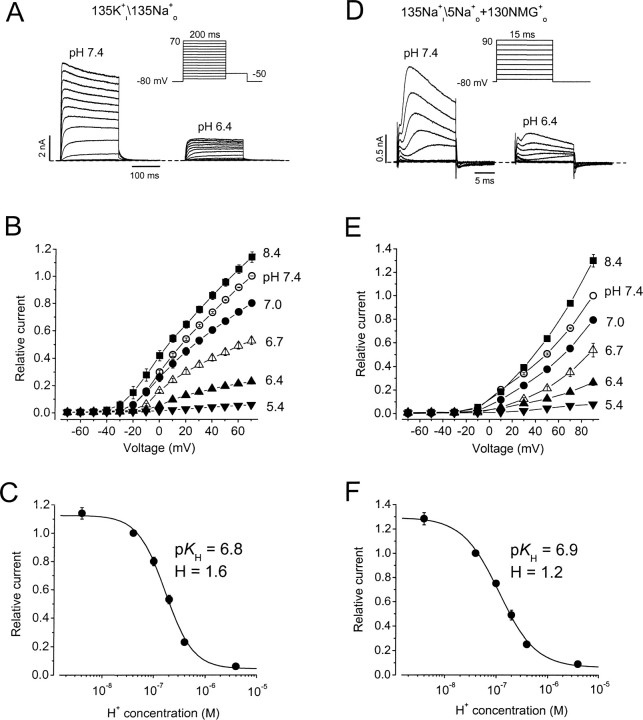
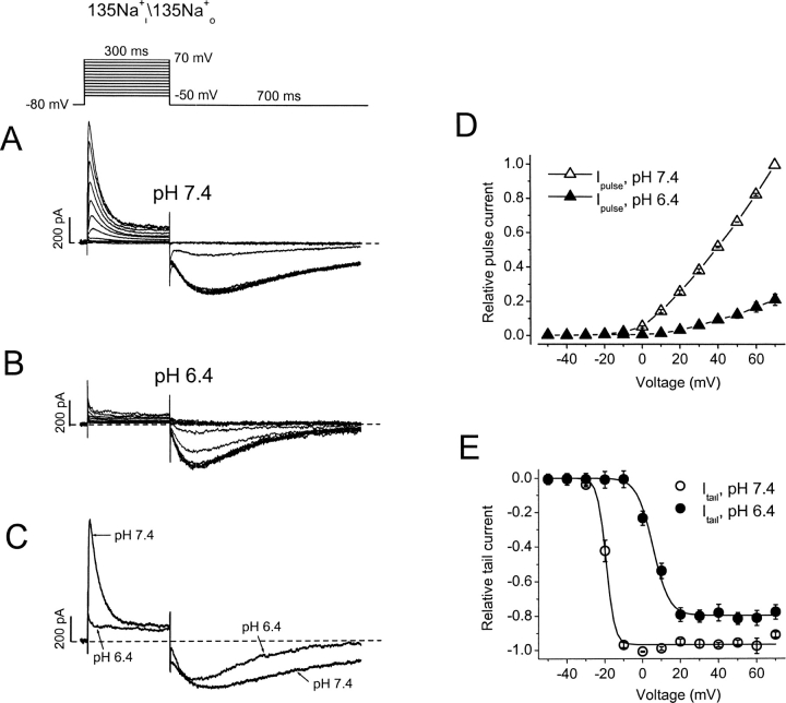
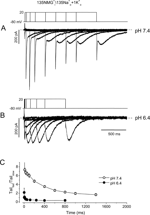
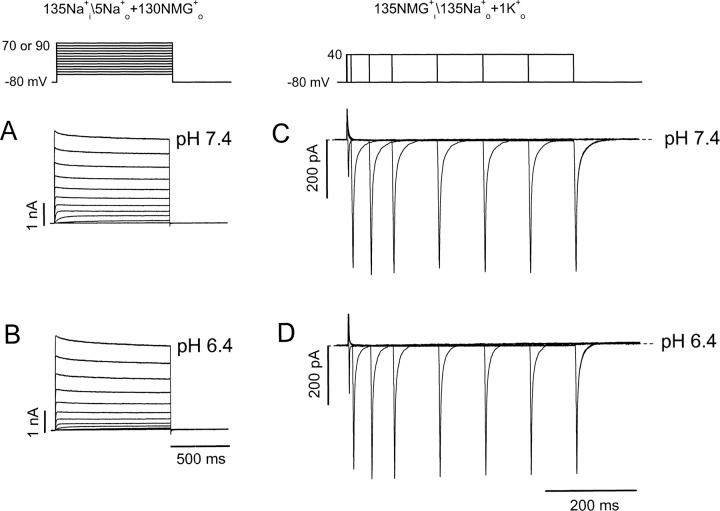
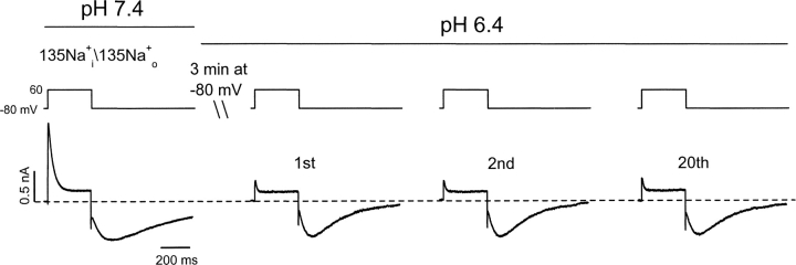
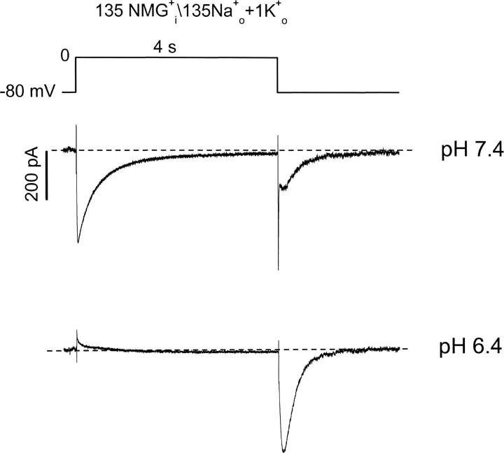
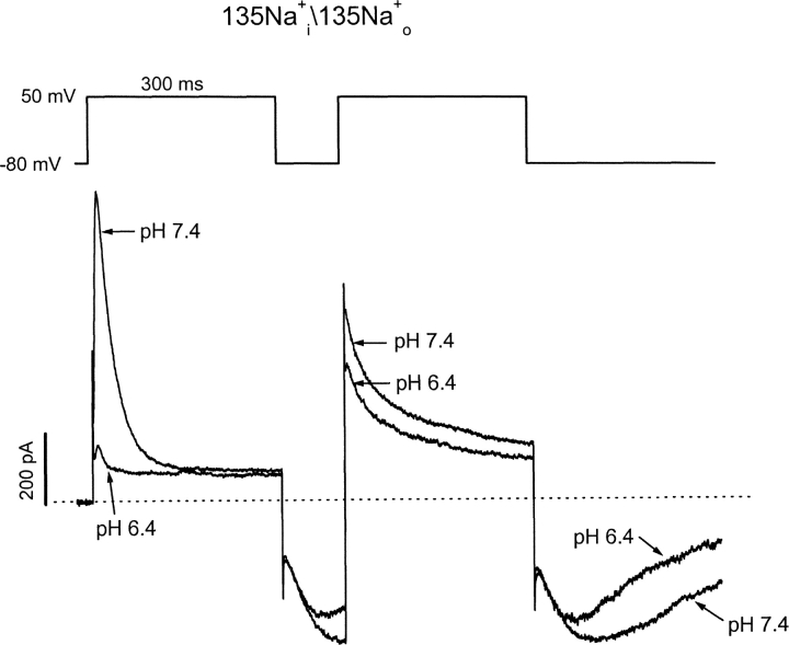
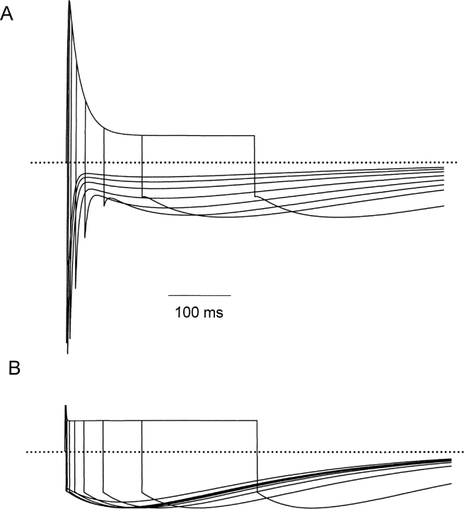
Extracellular acidification is known to decrease the conductance of many voltage-gated potassium channels. In the present study, we investigated the mechanism of H(+)(o)-induced current inhibition by taking advantage of Na(+) permeation through inactivated channels. In hKv1.5, H(+)(o) inhibited open-state Na(+) current with a similar potency to K(+) current, but had little effect on the amplitude of inactivated-state Na(+) current. In support of inactivation as the mechanism for the current reduction, Na(+) current through noninactivating hKv1.5-R487V channels was not affected by [H(+)(o)]. At pH 6.4, channels were maximally inactivated as soon as sufficient time was given to allow activation, which suggested two possibilities for the mechanism of action of H(+)(o). These were that inactivation of channels in early closed states occurred while hyperpolarized during exposure to acid pH (closed-state inactivation) and/or inactivation from the open state was greatly accelerated at low pH. The absence of outward Na(+) currents but the maintained presence of slow Na(+) tail currents, combined with changes in the Na(+) tail current time course at pH 6.4, led us to favor the hypothesis that a reduction in the activation energy for the inactivation transition from the open state underlies the inhibition of hKv1.5 Na(+) current at low pH.


Figures

F<sc>igure</sc> 1.
Figure 1.
Extracellular acidification inhibits hKv1.5 K+ and Na+ currents. (A–C) pH effects on K+ currents. A, hKv1.5 currents in response to a step voltage protocol at pH 7.4 or pH 6.4 from the same cell in nominally K+ o-free medium. Internal\external solution composition is shown above A and D. (B) Normalized mean peak I-V relationships at different pH from 3–5 cells. Data were normalized to peak outward current at 70 mV, pH 7.4. (C) Peak current amplitude at 70 mV normalized to the value at pH 7.4, and plotted versus pH. The K d was 166 nM, pK H was 6.8, and the Hill coefficient was 1.6. (D–F) As for A–C except that the pipette and bath solutions contained Na+ as the permeant ion, as indicated above D. In F the K d was 114 nM, pK H was 6.9, and the Hill coefficient was 1.2. No significant difference was detected between the values at the two pHs (P > 0.1). Lines through points in C and F are Hill fit lines, whereas those in B and E are to connect the points only.
F<sc>igure</sc> 2.
Figure 2.
Differential effects of extracellular acidification on Na+ currents through open versus inactivated hKv1.5 channels. (A and B) Na+ currents from the same cell in response to the step voltage protocol shown above the current traces at pH 7.4 and pH 6.4. Currents are shown for 10-mV steps. The interpulse interval was 10 s. (C) Comparison of current traces at 70 mV from A and B. (D and E) I-V relationships normalized to current at 70 mV and pH 7.4 for the peak outward current on depolarization and peak tail currents on repolarization. Note that acidification dramatically reduced the outward Na+ current without substantially affecting the tail currents. With extracellular acidification the peak current at 70 mV decreased by 78.8 ± 3.3% (n = 6, P < 0.01), whereas the maximal Na+ tail current was reduced by 18.5 ± 4.4% (n = 6, P < 0.01). Acidification caused a rightward shift of the voltage (V 1/2) for the half maximal Na+ tail current amplitude by 24.7 mV from −19.3 ± 0.4 to 5.4 ± 0.6 mV. Lines through points in E are Boltzmann fit lines, whereas those in D are to connect the points only.
F<sc>igure</sc> 3.
Figure 3.
Extracellular acidification promotes inactivation. (A and B) Superimposed tail currents after depolarizations of varying duration at pH 7.4 and pH 6.4. Pipette contained NMG+ as the major intracellular cation, and the bath solution contained 135 Na+ o + 1 mM K+ o. On the first pulse the membrane was depolarized from −80 to 20 mV for 2 ms at pH 7.4, and 1 ms at pH 6.4. For subsequent pulses the protocol was repeated every 10 s for increasing durations, 10, 30, 60, 100, 200, 400, 600, 800, 1,000, and 1,400 ms at pH 7.4; and 4, 8, 16, 24, 32, 40, 60, 120, 240, 400, and 800 ms at pH 6.4. Tail currents are composed of a fast component, reflecting deactivation from the open state, and a slow component reflecting Na+ current through inactivated channels. (C) The ratio of peak transient inward current amplitude versus the peak slow tail current is plotted against the duration of depolarization at 20 mV at pH 7.4 (○) and pH 6.4 (•).
F<sc>igure</sc> 4.
Figure 4.
Extracellular acidification does not inhibit Na+ current through the inactivation-deficient mutant hKv1.5-R487V. (A and B) Na+ currents from the same cell in response to the step voltage protocol shown above the current traces at pH 7.4 and pH 6.4. (C and D) Superimposed tail currents during the envelope of voltage steps shown above the current traces at pH 7.4. The depolarization to 40 mV was initially 1 ms in duration and the protocol was repeated every 10 s for increasing durations up to 500 ms.
F<sc>igure</sc> 5.
Figure 5.
Inactivation of Kv1.5 current is complete at the first depolarization in low pH. In symmetrical Na+ i/Na+ o solutions, control Na+ current was recorded by applying the voltage protocol shown above the current traces at pH 7.4. The pH of the bath solution was then changed to 6.4 while the cell was held at −80 mV for 3 min. Na+ currents at pH 6.4 were recorded again by applying the same voltage protocol every 10 s. The first depolarization elicited a response in which the outward current was largely eliminated but the tail current remained well developed. The waveform of the Na+ current remained unchanged over the next 20 pulses at pH 6.4 (n = 6).
F<sc>igure</sc> 6.
Figure 6.
Extracellular acidification causes early and complete Kv1.5 channel inactivation. Kv1.5 channel current was elicited by depolarization to 0 mV for 4 s at pH 7.4. The inclusion of 1 mM K+ o allowed an inactivating inward K+ current through the open state to be seen. Repolarization tail currents with fast (K+) and slow (Na+) components were also recorded. The pH of bath solution was then changed to 6.4 while the cell was held at −80 mV for 3 min. The current during the first depolarization of the same voltage protocol at pH 6.4 is shown in the bottom panel (n = 7).
F<sc>igure</sc> 7.
Figure 7.
Slowly inactivating outward currents upon activation from closed inactivated states. In symmetrical Na+ i/Na+ o solutions, Kv1.5 channel current was elicited by depolarization to 50 mV for 300 ms at pH 7.4. After a brief 100-ms repolarization, a second test pulse to 50 mV was given near the peak of the slow inward tail current. The pH of the bath solution was then changed to 6.4 while the cell was held at −80 mV for 3 min, and the two-step voltage protocol was repeated (n = 7). Note the much slower inactivation of R-state “supernormal” current during the second step pulse at both pH 7.4 and 6.4.
F<sc>igure</sc> 8.
Figure 8.
Kinetic model describing Na+ permeation through WT Kv1.5. (A) All rate constants in the activation pathway were governed by exponential functions of voltage (see materials and methods), and were taken from a previously published model of Kv1.5 inactivation (Kurata et al., 2001). At 0 mV, the rate constants were (in ms−1) kf = 0.4, kb = 0.065, ko = 0.31, and kob = 0.017, with associated valences of zf = 1.4, zb = −0.35, zo = 0.60, and zob = −0.70. Closed-state inactivation and recovery transitions were voltage independent, but were allosterically coupled to transitions in the activation pathway. Rate constants were (ms−1) kci = 0.00008, kic = 0.000055, and the allosteric factor f was 0.64. Rate constants in the recovery pathway to simulate currents at pH = 7.4 were (ms−1) kio = 0.00125, koi = 0.05, kri = 0.0072, kir = 0.000323, kcr = 0.185, krc = 0.00855. Transitions between I, R, and I4 were exponential functions of voltage with equivalent valences for zri= 0.55, zir = −0.65, zcr = 0.05, and zrc = −0.05. The relative conductances γ of the open, inactivated states and R states were 1.0, 0.1, and 1.0, respectively. (B) Rate kci was increased from 0.00008 ms−1 to 0.8 ms−1, to simulate channel inactivation occurring primarily from closed states. (C) Rates kio and koi were accelerated 50-fold to simulate the currents observed at pH = 6.4.
F<sc>igure</sc> 9.
Figure 9.
Simulation of the development of Na+ currents at pH 7.4 or 6.4. Simulations were performed using the parameters described in Fig. 8 C for pH 7.4 (A) and pH 6.4 (B). In A and B, currents were simulated with depolarizing stimuli to 60 mV for 2, 4, 8, 15, 30, 60, 120, and 300 ms, followed by a repolarization to −80 mV. The simulations illustrate the more rapid development of the Na+ tail at pH 6.4, and also clearly illustrate the rapid rising phase and rapid decay of the Na+ tail (compare Fig. 3).

Similar articles

Cited by

References

    1. Chen, F.S.P., D. Steele, and D. Fedida. 1997. Allosteric effects of permeating cations on gating currents during K+ channel deactivation. J. Gen. Physiol. 110:87–100. - PMC - PubMed
    1. Claydon, T.W., M.R. Boyett, A. Sivaprasadarao, K. Ishii, J.M. Owen, H.A. O'Beirne, R. Leach, K. Komukai, and C.H. Orchard. 2000. Inhibition of the K+ channel Kv1.4 by acidosis: protonation of an extracellular histidine slows the recovery from N-type inactivation. J. Physiol. 526:253–264. - PMC - PubMed
    1. Claydon, T.W., M.R. Boyett, A. Sivaprasadarao, and C.H. Orchard. 2002. Two pore residues mediate acidosis-induced enhancement of C-type inactivation of the Kv1.4 K+ channel. Am. J. Physiol. Cell Physiol. 283:C1114–C1121. - PubMed
    1. Eisner, D.A., C.G. Nichols, S.C. O'Neill, G.L. Smith, and M. Valdeolmillos. 1989. The effects of metabolic inhibition on intracellular calcium and pH in isolated rat ventricular cells. J. Physiol. 411:393–418. - PMC - PubMed
    1. Ishii, K., K. Nunoki, T. Yamagishi, H. Okada, and N. Taira. 2001. Differential sensitivity of Kv1.4, Kv1.2, and their tandem channel to acidic pH: involvement of a histidine residue in high sensitivity to acidic pH. J. Pharmacol. Exp. Ther. 296:405–411. - PubMed

Publication types