. 2003 Mar;121(3):227-44.
doi: 10.1085/jgp.20028768.

On the conformation of the COOH-terminal domain of the large mechanosensitive channel MscL

Affiliations

On the conformation of the COOH-terminal domain of the large mechanosensitive channel MscL

Andriy Anishkin et al. J Gen Physiol. 2003 Mar.

Abstract

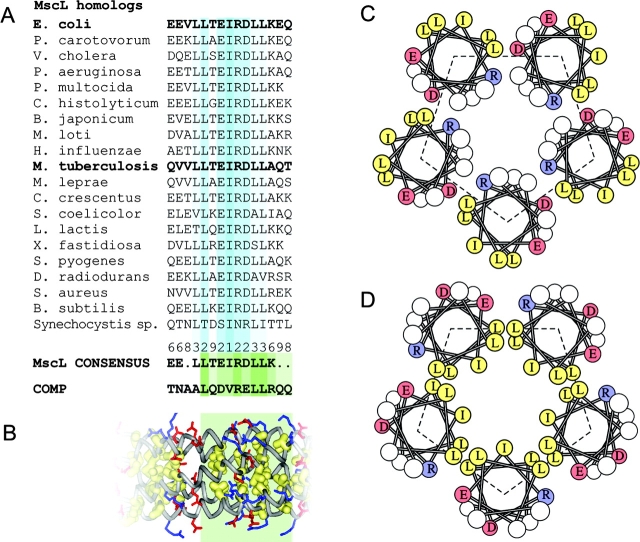
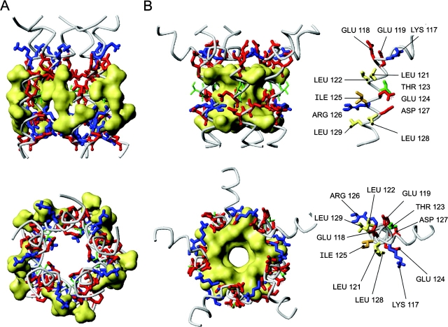
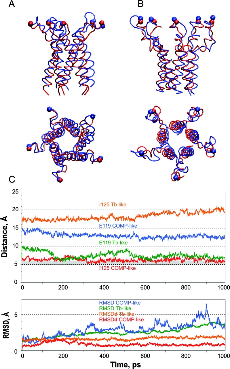
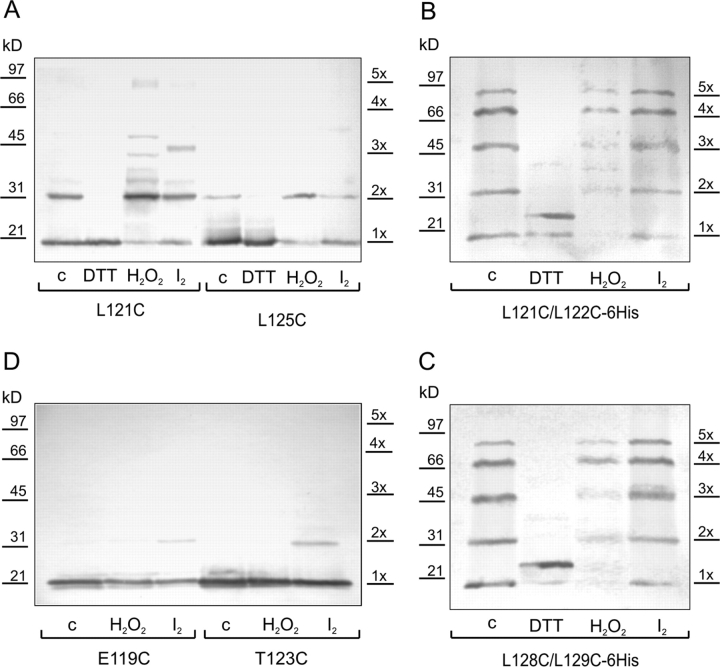
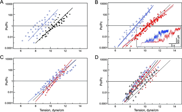
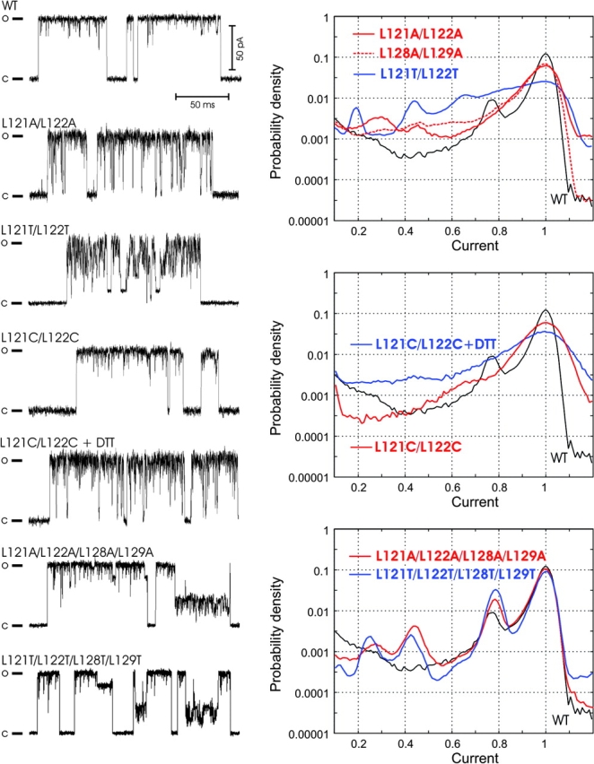
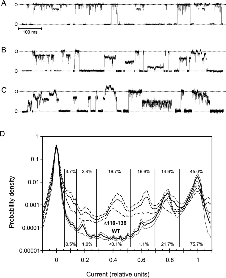
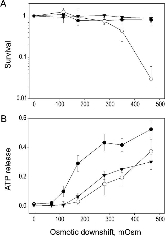
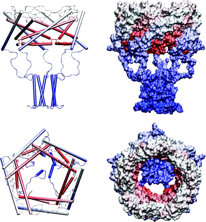
COOH-terminal (S3) domains are conserved within the MscL family of bacterial mechanosensitive channels, but their function remains unclear. The X-ray structure of MscL from Mycobacterium tuberculosis (TbMscL) revealed cytoplasmic domains forming a pentameric bundle (Chang, G., R.H. Spencer, A.T. Lee, M.T. Barclay, and D.C. Rees. 1998. SCIENCE: 282:2220-2226). The helices, however, have an unusual orientation in which hydrophobic sidechains face outside while charged residues face inside, possibly due to specific crystallization conditions. Based on the structure of pentameric cartilage protein, we modeled the COOH-terminal region of E. coli MscL to better satisfy the hydrophobicity criteria, with sidechains of conserved aliphatic residues all inside the bundle. Molecular dynamic simulations predicted higher stability for this conformation compared with one modeled after the crystal structure of TbMscL, and suggested distances for disulfide trapping experiments. The single cysteine mutants L121C and I125C formed dimers under ambient conditions and more so in the presence of an oxidant. The double-cysteine mutants, L121C/L122C and L128C/L129C, often cross-link into tetrameric and pentameric structures, consistent with the new model. Patch-clamp examination of these double mutants under moderately oxidizing or reducing conditions indicated that the bundle cross-linking neither prevents the channel from opening nor changes thermodynamic parameters of gating. Destabilization of the bundle by replacing conservative leucines with small polar residues, or complete removal of COOH-terminal domain (Delta110-136 mutation), increased the occupancy of subconducting states but did not change gating parameters substantially. The Delta110-136 truncation mutant was functional in in vivo osmotic shock assays; however, the amount of ATP released into the shock medium was considerably larger than in controls. The data strongly suggest that in contrast to previous gating models (Sukharev, S., M. Betanzos, C.S. Chiang, and H.R. Guy. 2001a. NATURE: 409:720-724.), S3 domains are stably associated in both closed and open conformations. The bundle-like assembly of cytoplasmic helices provides stability to the open conformation, and may function as a size-exclusion filter at the cytoplasmic entrance to the MscL pore, preventing loss of essential metabolites.


Figures

F<sc>igure</sc> 1.
Figure 1.
Sequence analysis of conserved COOH-terminal regions of MscL and comparison with the cartilage protein COMP. Alignment of S3 domains (residues 118–132 for EcoMscL) for 19 representative MscL homologues from different bacterial species, the consensus and the sequence of the third heptad of the oligomerization domain of COMP (A). The fragment of the crystal structure of COMP representing the aligned sequence (green box), which shows all hydrophobic side chains packed inside the fivefold coiled-coil and the salt bridges between R48 or R52 (blue) and D46 (red) on different chains (B). Helical wheel representations of the packing of helices in the crystal structure of TbMscL (C) and in EcoMscL modeled after COMP (D).
F<sc>igure</sc> 2.
Figure 2.
Models of the cytoplasmic bundle of EcoMscL. (A) A model built after the crystal structure of TbMscL as a left-handed bundle with sidechains of the conserved aliphatic residues L121, L122, I125, L128, and L129 all outside (shown by yellow surfaces). (B) A COMP-like model of S3 bundle. All five helices form a short left-handed coiled coil with a 19° tilt. All aliphatic side chains are inside, collectively shown by surface. Two rings of potential salt bridges were inferred. Positions of critical side chains on an individual S3 helix are shown on the right.
F<sc>igure</sc> 3.
Figure 3.
Molecular simulations of the COOH-terminal domains of EcoMscL. The results of 1-ns simulation of (A) Tb-like model, and (B) COMP-like model at 310 K. The initial conformations are represented as red tube; the final frames are shown as blue tube. (C) Distance trajectories for representative pairs of equivalent residues on the hydrophobic (I125) and polar (E119) sides of adjacent S3 helixes. The bottom panel shows root mean square deviations of absolute coordinates (RMSD) and of relative distances (RMSDd) derived from the 310K trajectories of Cβ atoms of the residues K106, P109, and A112 (linkers) and L121, I125, and L129 (bundle).
F<sc>igure</sc> 4.
Figure 4.
The virtual “pull” experiment in which Cα atoms of residues R104 (A) or residues A114 (B) were steered to new positions shown by arrows. A harmonic force with the constant of 1 kcal/mole*Å2 was applied to those atoms shown as balls. The initial positions are depicted in red; the final frames after 2 ns simulation are shown in blue. The bundle remained stable during both simulations.
F<sc>igure</sc> 5.
Figure 5.
Disulfide cross-linking in single and double cysteine MscL mutants. The patterns were visualized with the Western blot technique. Cross-linking conditions are denoted as c (control) - ambient oxygen, DTT (10 mM), H2O2 (0.3%), I2 (0.1 mM), duration of all treatments was 15 min.
F<sc>igure</sc> 6.
Figure 6.
Dose–response curves for WT MscL, S3 mutants with substituted leucines, and the Δ110–136 truncation mutant. The data is presented as Po/Pc values versus membrane tension. Four to seven curves measured on independent patches were plotted and fitted according to the two-state model (see materials and methods). The lines represent curves with average parameters ΔE and ΔA (see Table III) for each dataset. (A) WT MscL measured under ambient conditions (filled circles, black solid line) or in the presence of 25 mM DTT (blue open circles, dotted line). (B) L121C/L122C (squares, solid line) and L128C/L129C (triangles, dotted line) measured under ambient (red) or reducing (blue) conditions. (set) Single-channel traces of L128C/L129C MscL measured under ambient conditions (red) or in the presence of 25 mM DTT (blue) in the same patch, in response to a similar sequence of pressure steps. (C) L121A/L122A (red) and L121T/L122T (blue) measured under ambient conditions. (D) The data for the quadruple L121A/L122A/L128A/L129A (red circles and solid line) and L121T/L122T/L128T/L129T (blue diamonds and dotted line) mutants and for the truncated Δ110–136 MscL mutant (triangles, black dashed line). The black solid line representing WT MscL is given as reference in each panel.
F<sc>igure</sc> 7.
Figure 7.
Representative single-channel traces (left) and probability density amplitude histograms (right) for WT MscL and mutants with alanines, threonines, or cysteines substituting for conserved leucines in S3. Cysteine mutants were characterized under ambient or reducing conditions. The histograms for WT are shown in all panels as reference (black solid line).
F<sc>igure</sc> 8.
Figure 8.
Fragments of typical patch-clamp traces recorded from giant spheroplasts expressing Δ110–136 MscL. The mutant shows WT-like gating patterns (A), but greater variability of single-channel amplitudes and kinetic patterns (B and C). (D) Probability density distributions of current amplitudes for Δ110–136 and WT calculated from all-point histograms. To emphasize only long-lived subconducting states, fragments of the traces were low-pass filtered at a 500 Hz cut-off. The fragments of traces characterized with Po between 0.4 and 0.5 were selected. The current was normalized to the amplitude of the fully open state and the range from −0.1 to 1.1 was divided into 120 bins. Thick lines represent the mean of 11 traces for WT and 10 traces for Δ110–136. Thin lines delineate standard error intervals calculated for each bin. The relative occupancy for each substate indicated by numbers was calculated as the fraction of data points between the two minimums separating the adjacent peaks.
F<sc>igure</sc> 9.
Figure 9.
The survival of MJF 455 E. coli cells (A) and ATP release (B) under conditions of varied osmotic shock. The cells were grown in a rich medium of 850 mOsm and subjected to osmotic downshifts of indicated magnitudes. The ATP in the shock medium was assayed, and the same samples were plated for colony count. The empty vector control is shown by open circles, WT-expresser cells by triangles, and Δ110–136 cells by filled circles. The data represent means of three independent experiments (± SEM).
F<sc>igure</sc> 10.
Figure 10.
Structural models of EcoMscL in the open conformation. The right side shows α helices as cylinders and coiled segments as strings, the left side is a space-filled model. The structure begins with red at the NH2 terminus and ends with blue at the COOH terminus. The top figures show a side view and the bottom shows a view from the outside through the pore. The five windows of the sieve are formed by the linkers that connect the M2 transmembrane segments to the S3 helices.

References

    1. Ajouz, B., C. Berrier, A. Garrigues, M. Besnard, and A. Ghazi. 1998. Release of thioredoxin via the mechanosensitive channel MscL during osmotic downshock of Escherichia coli cells. J. Biol. Chem. 273:26670–26674. - PubMed
    1. Arkin, I.T., S.I. Sukharev, P. Blount, C. Kung, and A.T. Brunger. 1998. Helicity, membrane incorporation, orientation and thermal stability of the large conductance mechanosensitive ion channel from E. coli. Biochim. Biophys. Acta. 1369:131–140. - PubMed
    1. Betanzos, M., C.-S. Chiang, H.R. Guy, and S. Sukharev. 2002. A large iris-like expansion of a mechanosensitive channel protein induced by membrane tension. Nat. Struct. Biol. 9:704. - PubMed
    1. Bian, S., I. Favre, and E. Moczydlowski. 2001. Ca2+-binding activity of a COOH-terminal fragment of the Drosophila BK channel involved in Ca2+-dependent activation. Proc. Natl. Acad. Sci. USA. 98:4776–4781. - PMC - PubMed
    1. Blount, P., S.I. Sukharev, P.C. Moe, M.J. Schroeder, H.R. Guy, and C. Kung. 1996. a. Membrane topology and multimeric structure of a mechanosensitive channel protein of Escherichia coli. EMBO J. 15:4798–4805. - PMC - PubMed

Publication types

MeSH terms