. 2003 Mar 4;100(5):2278-83.
doi: 10.1073/pnas.0537525100. Epub 2003 Feb 24.

The ATP hydrolyzing transcription activator phage shock protein F of Escherichia coli: identifying a surface that binds sigma 54

Affiliations

The ATP hydrolyzing transcription activator phage shock protein F of Escherichia coli: identifying a surface that binds sigma 54

Patricia Bordes et al. Proc Natl Acad Sci U S A. .

Abstract

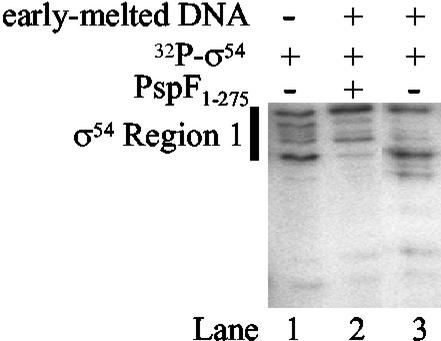
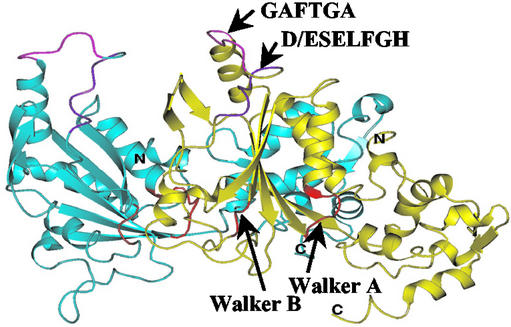
Members of the protein family called ATPases associated with various cellular activities (AAA(+)) play a crucial role in transforming chemical energy into biological events. AAA(+) proteins are complex molecular machines and typically form ring-shaped oligomeric complexes that are crucial for ATPase activity and mechanism of action. The Escherichia coli transcription activator phage shock protein F (PspF) is an AAA(+) mechanochemical enzyme that functions to sense and relay the energy derived from nucleoside triphosphate hydrolysis to catalyze transcription by the sigma(54)-RNA polymerase. Closed promoter complexes formed by the sigma(54)-RNA polymerase are substrates for the action of PspF. By using a protein fragmentation approach, we identify here at least one sigma(54)-binding surface in the PspF AAA(+) domain. Results suggest that ATP hydrolysis by PspF is coupled to the exposure of at least one sigma(54)-binding surface. This nucleotide hydrolysis-dependent presentation of a substrate binding surface can explain why complexes that form between sigma(54) and PspF are transient and could be part of a mechanism used generally by other AAA(+) proteins to regulate activity.


Figures

Figure 1
Figure 1
Schematic showing the domain organization of E. coli PspF (to scale). PspF contains 325 amino acid residues and is composed of two domains. The carboxyl-terminal domain (residues 296–325, highlighted in black) contains a helix-turn-helix DNA-binding domain. (Top) The amino-terminal AAA+ domain (residues 1–275) is directly responsible for ATP hydrolysis and transcriptional activation. This domain is highly conserved among σ54 activators; the seven conserved regions (C1–C7) are indicated in gray. The Walker A and Walker B motifs involved in NTP binding and hydrolysis are indicated. The amino acid sequences of the PspF fragments F1 (residues 69–134), F2 (residues 69–102), F3 (residues 89–134), and F4 (residues 69–93) are shown. (Middle) C3 region containing the signature motif of σ54 activators, D/ESELFGH and GAFTGA, is shown in box. (Bottom) Walker B motif is boxed. Residues that were targeted for mutagenesis are highlighted (see text).
Figure 2
Figure 2
Stable binding of PspF fragment MBP-F4 to a σ54–early-melted promoter probe complex. (A) Sequences (residues shown are −35 to +3) of the S. meliloti homoduplex and early-melted promoter probes. (B) Native gel showing the NTP-independent binding of PspF MBP-F4 to early-melted promoter probe bound σ54. The background smear migrating at the same position as the σ54–early-melted probe complex is present in all lanes, even if protein is added. (C) Native gel showing that σ54 is part of the PspF MBP-F4-dependent complex. Reactions were carried out as in B but, where indicated, contained either 16 nM 32P-end-labeled early-melted DNA (lanes 1–3) or 1 μM 32P-end-labeled σ54 (lanes 4–5). (D) Western blot analysis of the protein content of the PspF MBP-F4-dependent complex. (a) Native gel showing PspF MBP-F4 complex formation. Proteins in the boxed complexes were blotted by using antibodies against the anti-RNAP β-subunit (b) and E. coli anti-MBP protein (c). Lanes 1 and 2 contain purified E. coli core RNAP and PspF MBP-F4, respectively. Lanes 3 and 4 contain the contents of the gel-isolated complexes C1 and C2, respectively. Arrows indicate the migration position of the E. coli RNAP and purified PspF MBP-F4.
Figure 3
Figure 3
Region 1 of σ54 and the GAFTGA motif of PspF are required for stable formation of the PspF MBP-F4-σ54–early-melted DNA complex. (A) Native gel showing the reduced ability of PspF MBP-F4 to form a stable complex with ΔR1σ54 bound to the early-melted promoter probe. For the reaction shown in lane 4, σ54 region 1 was added in trans to the ΔR1σ54–early-melted promoter probe complex before adding PspF F4. (B) Native gel showing the ability of mutant PspF MBP-F4 fragments to form complexes with early-melted bound σ54.
Figure 4
Figure 4
DNase I footprint of the PspF MBP-F4 complex. (A) Native gel showing PspF F4 complex formation. Unbound probe and complexes that were isolated for analysis by denaturing PAGE are boxed. (B) Denaturing gel (10%) showing σ54 and PspF MBP-F4 complex-dependent footprints on the early-melted probe. Solid line represents the DNA segment that becomes protected upon σ54 binding and hypersensitive upon PspF MBP-F4 complex formation. (C) Scan of lanes 4 and 5 from B. *, Positions in the promoter that become protected only in the PspF MBP-F4 complex. ↓, The enhanced reactivity of the DNA to DNase I around position −12 in the PspF MBP-F4 complex.
Figure 5
Figure 5
Analysis of PspF–σ54 interactions by V8 protease footprinting. Lane 1, V8 protease digestion pattern of 32P-σ54; lane 3, when bound to the early-melted promoter probe; lane 2, V8 digestion of 32P-σ54 bound to the early-melted promoter probe in the presence of unlabeled PspF1–275. Region 1 of σ54 is indicated by a thick black line.
Figure 6
Figure 6
Homology model of the PspF dimer based on the crystal structures of HslU and p97 (5). One promoter is colored blue and the other is colored yellow. The GAFTGA (in pink) and the D/ESELFGH (in purple) motifs are indicated. The Walker A and Walker B motifs implicated in NTP interactions are shown in red.

References

    1. Neuwald A F, Aravind L, Spouge J L, Koonin E V. Genome Res. 1999;9:27–43. - PubMed
    1. Vale R D. J Cell Biol. 2000;150:13–19. - PMC - PubMed
    1. Buck M, Gallegos M T, Studholme D J, Guo Y, Gralla J D. J Bacteriol. 2000;182:4129–4136. - PMC - PubMed
    1. Xu H, Hoover T R. Curr Opin Microbiol. 2001;4:138–144. - PubMed
    1. Zhang X, Chaney M K, Wigneshweraraj S R, Schumacher J, Bordes P, Cannon W, Buck M. Mol Microbiol. 2002;45:895–903. - PubMed

Publication types

MeSH terms

LinkOut - more resources