Oestrogen receptor specificity in oestradiol-mediated effects on B lymphopoiesis and immunoglobulin production in male mice
- PMID: 12603601
- PMCID: PMC1782901
- DOI: 10.1046/j.1365-2567.2003.01599.x
Oestrogen receptor specificity in oestradiol-mediated effects on B lymphopoiesis and immunoglobulin production in male mice
Abstract
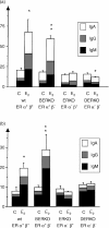
Oestrogen treatment down-regulates B lymphopoiesis in the bone marrow of mice. Meanwhile it up-regulates immunoglobulin production. To understand better the oestrogen action on bone marrow male mice lacking oestrogen receptor alpha (ERalpha; ERKO mice), lacking ERbeta (BERKO mice), lacking both receptors (DERKO mice) or wild-type (wt) littermates were castrated and treated for 2.5 weeks with 30 microg/kg 17beta-oestradiol (E2) or vehicle oil as controls. The B lymphopoiesis in the bone marrow was examined by flow cytometry and mature B-cell function was studied using an ELISPOT assay enumerating the B cells in bone marrow and spleen that were actively producing immunoglobulins. In wt mice the frequency of B-lymphopoietic (B220+) cells in the bone marrow decreased from 15% to 5% upon E2 treatment. In ERKO and BERKO mice significant reduction was seen but not of the same magnitude. In DERKO mice no reduction of B lymphopoiesis was seen. In addition, our results show that E2 mediated reduction of different steps in B lymphopoiesis require only ERalpha or both receptors. In wt and BERKO mice E2 treatment resulted in significantly increased levels of B cells actively producing immunoglobulin, while in ERKO and DERKO mice no such change was seen. Similar results were found in both bone marrow and spleen. In conclusion our results clearly show that both ERalpha and ERbeta are required for complete down-regulation of B lymphopoiesis while only ERalpha is needed to up-regulate immunoglobulin production in both bone marrow and spleen.
Figures


Similar articles
-
Influence of oestrogen receptor alpha and beta on the immune system in aged female mice.Immunology. 2003 Sep;110(1):149-57. doi: 10.1046/j.1365-2567.2003.01704.x. Immunology. 2003. PMID: 12941152 Free PMC article.
-
Effects of a tissue-selective estrogen complex on B lymphopoiesis and B cell function.Immunobiology. 2017 Aug;222(8-9):918-923. doi: 10.1016/j.imbio.2017.05.013. Epub 2017 May 16. Immunobiology. 2017. PMID: 28551078
-
Estrogenic agonism and antagonism of the soy isoflavone genistein in uterus, bone and lymphopoiesis in mice.APMIS. 2005 May;113(5):317-23. doi: 10.1111/j.1600-0463.2005.apm_113502.x. APMIS. 2005. PMID: 16011657
-
Estrogen receptors in skeletal metabolism: lessons from genetically modified models of receptor function.Crit Rev Eukaryot Gene Expr. 2002;12(2):89-100. doi: 10.1615/critreveukaryotgeneexpr.v12.i2.10. Crit Rev Eukaryot Gene Expr. 2002. PMID: 12434924 Review.
-
Regulation of murine B lymphopoiesis by stromal cells.Immunol Rev. 2021 Jul;302(1):47-67. doi: 10.1111/imr.12973. Epub 2021 May 17. Immunol Rev. 2021. PMID: 34002391 Review.
Cited by
-
Hormones and B-cell development in health and autoimmunity.Front Immunol. 2024 Apr 12;15:1385501. doi: 10.3389/fimmu.2024.1385501. eCollection 2024. Front Immunol. 2024. PMID: 38680484 Free PMC article. Review.
-
Sex Hormones and Lung Inflammation.Adv Exp Med Biol. 2021;1304:259-321. doi: 10.1007/978-3-030-68748-9_15. Adv Exp Med Biol. 2021. PMID: 34019274
-
The Role of Inflammatory Cytokines, the RANKL/OPG Axis, and the Immunoskeletal Interface in Physiological Bone Turnover and Osteoporosis.Scientifica (Cairo). 2013;2013:125705. doi: 10.1155/2013/125705. Epub 2013 Feb 3. Scientifica (Cairo). 2013. PMID: 24278766 Free PMC article. Review.
-
Influence of oestrogen receptor alpha and beta on the immune system in aged female mice.Immunology. 2003 Sep;110(1):149-57. doi: 10.1046/j.1365-2567.2003.01704.x. Immunology. 2003. PMID: 12941152 Free PMC article.
-
Immune dynamics throughout life in relation to sex hormones and perspectives gained from gender-affirming hormone therapy.Front Immunol. 2025 Jan 16;15:1501364. doi: 10.3389/fimmu.2024.1501364. eCollection 2024. Front Immunol. 2025. PMID: 39885993 Free PMC article. Review.
References
-
- Medina KL, Strasser A, Kincade PW. Estrogen influences the differentiation, proliferation, and survival of early B-lineage precursors. Blood. 2000;95:2059–67. - PubMed
-
- Rijhsinghani AG, Thompson K, Bhatia SK, Waldschmidt TJ. Estrogen blocks early T cell development in the thymus. Am J Reprod Immunol. 1996;36:269–77. - PubMed
-
- Josefsson E, Tarkowski A, Carlsten H. Anti-inflammatory properties of estrogen. I. In vivo suppression of leukocyte production in bone marrow and redistribution of peripheral blood neutrophils. Cell Immunol. 1992;142:67–78. - PubMed
-
- Carlsten H, Verdrengh M, Taube M. Additive effects of suboptimal doses of estrogen and cortisone on the suppression of T lymphocyte dependent inflammatory responses in mice. Inflamm Res. 1996;45:26–30. - PubMed
-
- Hanna N, Schneider M. Enhancement of tumor metastasis and suppression of natural killer cell activity by beta-estradiol treatment. J Immunol. 1983;130:974–80. - PubMed
Publication types
MeSH terms
Substances
LinkOut - more resources
Full Text Sources
Molecular Biology Databases
Research Materials

