Comparative Study
. 2003 Mar;47(5):1207-24.
doi: 10.1046/j.1365-2958.2003.03366.x.

Iron and contact with host cells induce expression of adhesins on surface of Trichomonas vaginalis

Affiliations
Comparative Study

Iron and contact with host cells induce expression of adhesins on surface of Trichomonas vaginalis

Ana F Garcia et al. Mol Microbiol. 2003 Mar.

Abstract

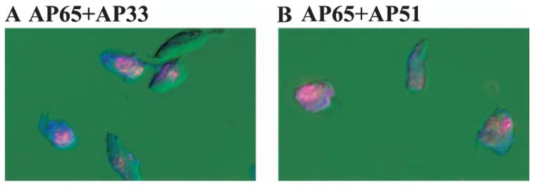
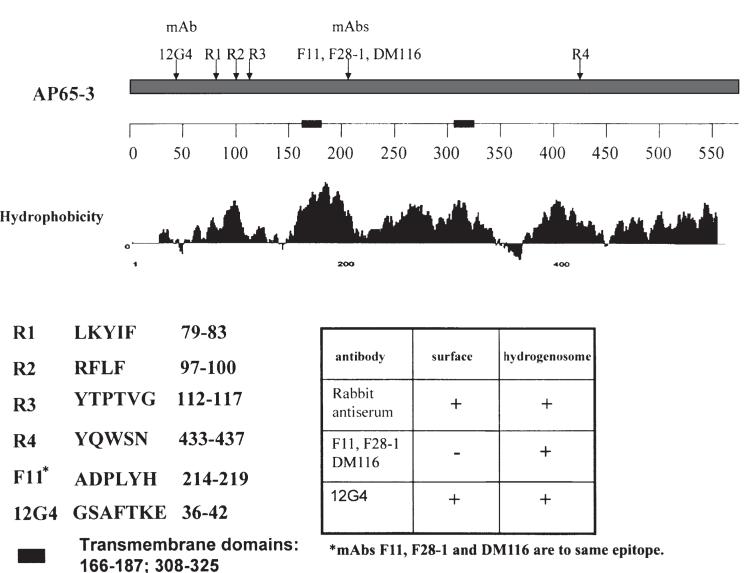
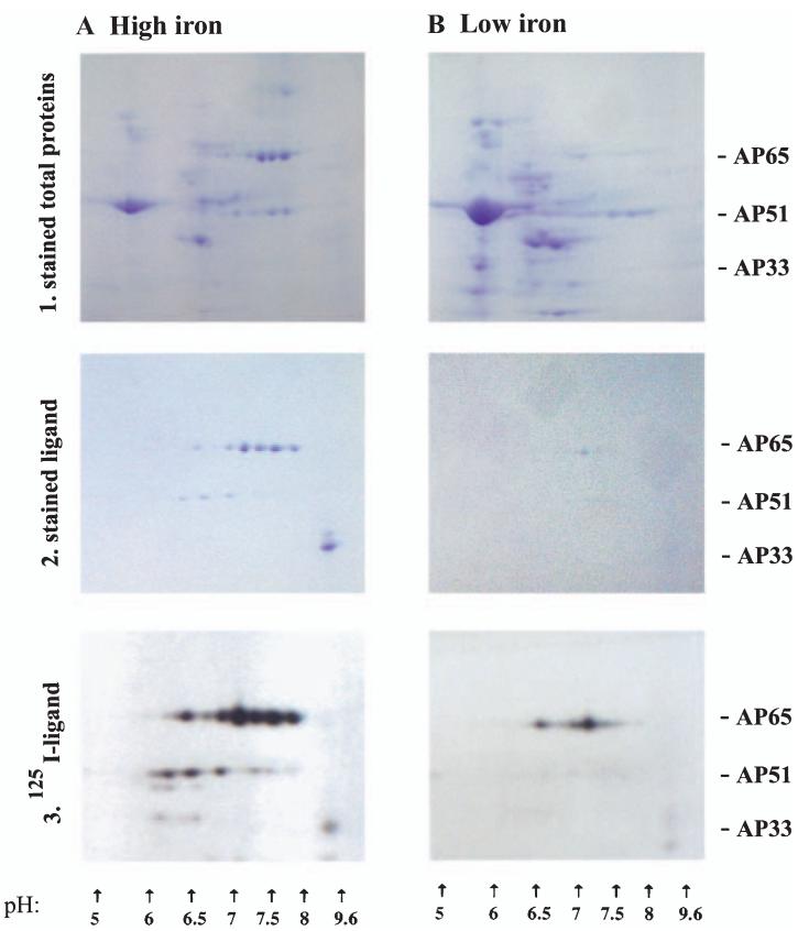
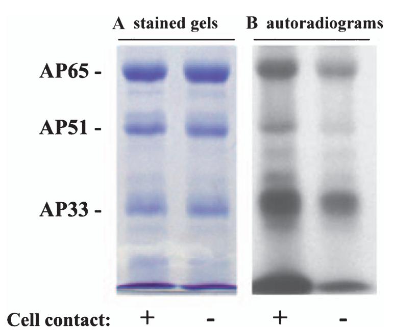
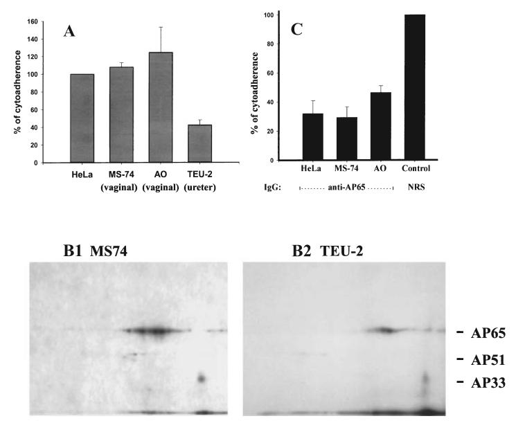
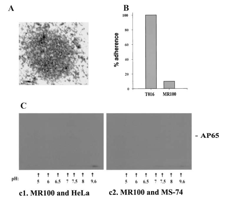
The proteins AP65, AP51, AP33 and AP23 synthesized by Trichomonas vaginalis organisms in high iron play a role in adherence. Multigene families encode enzymes of the hydrogenosome organelles, which have identity to adhesins. This fact raises questions regarding the compartmentalization of the proteins outside the organelle and about the interactions of adhesins with host cells. Data here demonstrate the presence of the proteins outside the organelle under high-iron conditions. Fluorescence and immuno-cytochemical experiments show that high-iron-grown organisms coexpressed adhesins on the surface and intracellularly in contrast with low-iron parasites. Furthermore, the AP65 epitopes seen by rabbit anti-AP65 serum that blocks adherence and detects surface proteins were identified, and a mAb reacting to those epitopes recognized the trichomonal surface. Two-dimensional electrophoresis and immunoblot of adhesins from surface-labelled parasites provided evidence that all members of the multigene family were co-ordinately expressed and placed on the trichomonal surface. Similar two-dimensional analysis of proteins from purified hydrogenosomes obtained from iodinated trichomonads confirmed the specific surface labelling of proteins. Contact of trichomonads with vaginal epithelial cells increased the amount of surface-expressed adhesins. Moreover, we found a direct relationship between the levels of adherence and amount of adhesins bound to immortalized vaginal and ureter epithelial cells, further reinforcing specific associations. Finally, trichomonads of MR100, a drug-resistant isolate absent in hydrogenosome proteins and adhesins, were non-adherent. Overall, the results confirm an important role for iron and contact in the surface expression of adhesins of T. vaginalis organisms.


Figures

Fig. 1
Fig. 1
Demonstrations of surface colocalization of AP65 with either AP33 (A) or AP51 (B) by fluorescence using non-permeabilized trichomonads of isolate T016 grown in a high-iron medium as described in Experimental procedures. Organisms were treated with antiserum IgG to adhesins conjugated individually to fluorescein (anti-AP65) or rhodamine (both anti-AP51 and AP33). Use of the individual antisera gave only the respective colour, and blending was observed indicative of coexpression. Little or no fluorescence was observed with prebleed NRS or with trichomonads grown in a low-iron medium (Lehker et al., 1991; Arroyo et al., 1992). In this experiment, fluorescence was examined with an Axiophot II Zeiss epifluorescent microscope.
Fig. 2
Fig. 2
Immuno-electron microscopic detection of AP65 on cryosections on the surface and different sites of high-iron (A1) vs. low-iron grown (B1) trichomonads of Trichomonas vaginalis T068-II. Anti-AP65 serum IgG followed by gold-conjugated goat anti-rabbit IgG was used in this experiment. Large numbers of gold particles were evident within hydrogenosomes of high-iron (A2) parasites. Few particles (arrow) were detectable in the hydrogenosome (H) of low-iron (B2) organisms. Other antisera to adhesins gave similar results. The mAbs F11, F28-1 and DM116 (Fig. 3) to AP65 gave results like those in a2 and b2 in high- and low-iron respectively.
Fig. 3
Fig. 3
Identification of epitopes using overlapping decapeptides obtained for AP65-3 as described in the Experimental procedures section. The IgG1 mAb F11 and two new IgG1 mAbs, F28-1, and DM116 generated from distinct mAb libraries recognized the same epitope. A LamB-AP65 fusion protein used for immunization of mice resulted in generation of mAb 12G4 toward an epitope at the amino terminus of the clustered rabbit epitopes. The sequences of the epitopes are included on the left of the insert table, which presents the combined fluorescence and immunogold labelling results of separate experiments. The new mAb reacted with the parasite surface as shown in Fig. 4. Two transmembrane domains are represented on the numbered scale by bold, solid bars, and the amino acid numbers are indicated.
Fig. 4
Fig. 4
Fluorescence experiments showing detection of AP65 on the surface of non-permeabilized (A) and intracellularly of permeabilized (B) trichomonads of isolate T016 with mAb 12G4 characterized in Fig. 3. The mAbs F11 and L64 to AP65 and to an intracellular protein (Alderete et al., 1987), respectively, were immunoreactive only with permeabilized organisms (B3 and B5). No signal was evident with non-permeabilized trichomonads (A3 and A5). Brightfield photomicrographs of the same fields accompany the fluorescence pictures.
Fig. 5
Fig. 5
Combined ligand assay with two-dimensional SDS-PAGE confirms the surface residence of the adhesin family members. Isoelectric focusing separated the total proteins or proteins from the ligand assay in a pH 3-10 ampholine gradient followed by electrophoreses and staining or autoradiography. Part 1. Coomassie brilliant blue stained total protein patterns of the duplicate samples of surface-iodinated organisms used in Parts 2 and 3 below. Trichomonads were grown in high- (A) and low-iron medium (B). The complex patterns of proteins are representative of equivalent cell numbers treated identically, and the different patterns are indicative of the iron status of trichomonads. Part 2. Stained patterns of proteins following the ligand assay, which enriches for the host cell-binding proteins of extracts of parasites grown in high (A) and low-iron medium (B). The same number of cell equivalents was used in the ligand assays. Part 3. Autoradiogram patterns of the stained gels in Part 2 showing iodinated protein spots of the two-dimensional gels. The X-ray film was exposed 8 h for both samples handled identically. It is noteworthy that immunoblots of duplicate samples of the stained gels of part 2A were performed comparing the rabbit antisera to the adhesins with mAbs F11 (AP65) and F5 (AP33). Not unexpectedly based on results recently published for AP33 (Engbring and Alderete, 1998a), the patterns of antibody reactivities were identical with those seen here for AP65 and AP33.
Fig. 6
Fig. 6
Comparison of two-dimensional patterns of total proteins (A) versus hydrogenosome proteins (B) of surface-iodinated T. vaginalis used in the ligand assay. Immunoblots of proteins after the ligand assay probed with mAbs F11 (AP65) and F5 (AP33) show quantitatively greater amounts of proteins in the total protein extracts (A1) compared with hydrogenosome extracts (B1). Inserts on bottom left of A1 and B1 are the Coomassie brilliant blue stained two-dimensional gels. The multiple bands for AP65 and AP51 are readily visible. The spot with a high pI at the lower right side corresponds to AP33, as seen in Fig. 5. Parts A2 and B2 present autoradiograms of gels of iodinated proteins of total trichomonad and hydrogenosome (B2) extracts, respectively, used in the ligand assay. No labelled proteins were evident for the hydrogenosome two-dimensional patterns even with prolonged exposure to X-ray film.
Fig. 7
Fig. 7
Contact with host cells shows increased amounts of surface-expressed adhesins. In this experiment, high iron trichomonads were first incubated with monolayers of immortalized AO VECs before removing and iodinating (see Experimental procedures). Part A shows the same amount of proteins enriched by the ligand assay, as evidenced by the equal intensity of Coomassie brilliant blue stained gels. Autoradiograms in part B illustrate a greater intensity of signal of the surface-labelled proteins of trichomonads after contact with the host cells. Equivalent parasite numbers were used in both samples, which were handled identically. Identical results were obtained with MS74 VECs.
Fig. 8
Fig. 8
Associations between levels of adherence of T. vaginalis T016 with amounts of adhesins bound to host cell surfaces. Part A, a cytoadherence assay was performed with quadruplicate samples comparing HeLa cells with immortalized VECs (MS74 and AO) and a immortalized ureter epithelial cell line (TEU-2). Overnight cultures of the confluent monolayers of cells were incubated with equal numbers of highly motile and radiolabelled mid-logarithmic phase trichomonads (see Experimental procedures). All samples were handled identically. The adherence experiment was performed on five different occasions and with similar results. The mean of the results for each cell type was compared with that for HeLa cells, which were normalized to be 100%. Approximately 40% of parasites in the suspension were adherent for HeLa cells. Part B. Autoradiogram two-dimensional patterns showing the amounts of iodinated adhesins from an extract of surface-labelled parasites bound to the cell surfaces by the ligand assay. MS74 (B1), HeLa cells and AO VECs had increased amounts of bound adhesins compared with the TEU-2 ureter cells (B2). In this experiment, the two-dimensional ligand assay was performed identically using duplicate samples of the radioiodinated trichomonad proteins and fixed host cells. X-ray film was exposed for an identical period of time to the acrylamide gels. Part C. Inhibition of adherence was demonstrated with purified anti-AP65 IgG eluted from preparative blots of AP65 (Arroyo et al., 1993). Washed radiolabelled organisms were first pretreated with antibody before addition to the cell monolayers. The level of adherence achieved for control prebleed normal rabbit serum (NRS) IgG was similar to that obtained in a normal adherence assay using HeLa cells, and this was normalized to 100%.
Fig. 9
Fig. 9
Absence of detectable adhesins and non-adherence of T. vaginalis MR100. Part A. A representative picture showing minimal labelling with gold-conjugated anti-rabbit IgG following treatment of the preparation with IRS to anti-AP65 IgG, as seen in Fig. 2A(2). The absence of the well defined double membrane for hydrogenosomes was apparent. Similar results were seen regardless of the iron status of trichomonads. Part B. A representative adherence assay using AO VECs comparing MR100 with wild-type trichomonads of T. vaginalis isolate T016 (Arroyo et al., 1993). All experiments were in quadruplicate and performed at least five different times. Part C. Autoradiograms showing the lack of any radioiodinated proteins by the two-dimensional ligand assay. This two-dimensional -ligand assay was performed at the same time as the adherence experiment in Fig. 8C. Identical duplicate samples of the radioiodinated MR100 trichomonad proteins were mixed with fixed host cells. Also as above, the X-ray film was exposed for a period of time identical to the positive ligand assay of Fig. 8B(1). Only did prolonged exposure of the X-ray films over several days were some protein spots visualized that migrated in the same area of the adhesins.

Similar articles

Cited by

References

    1. Addis MF, Rappelli P, Cappuccinelli P, Fiori PL. Extracellular release by Trichomonas vaginalis of a NADP-dependent malic enzyme involved in pathogenicity. Microbial Pathogen. 1997;23:55–61. - PubMed
    1. Addis MF, Rappelli P, Fiori PL. Host and tissue specificity of Trichomonas vaginalis is not mediated by its known adhesion proteins. Infect Immun. 2000;68:4358–4360. - PMC - PubMed
    1. Alderete JF. Identification of immunogenic and antibody-binding proteins on the membrane of pathogenic Trichomonas vaginalis. Infect Immun. 1983;40:284–291. - PMC - PubMed
    1. Alderete JF. Enzyme-linked immunosorbent assay for detection of antibody to Trichomonas vaginalis: use of whole cells and aqueous extract as antigen. Br J Vener Dis. 1984;60:164–170. - PMC - PubMed
    1. Alderete JF. Iron modulates phenotypic variation and phosphorylation of P270 in double-stranded RNA virus-infected Trichomonas vaginalis. Infect Immun. 1999;67:4298–4302. - PMC - PubMed

Publication types

MeSH terms

Associated data