. 2003 Mar 3;160(5):645-55.
doi: 10.1083/jcb.200209023. Epub 2003 Feb 25.

A role of topoisomerase II in linking DNA replication to chromosome condensation

Affiliations

A role of topoisomerase II in linking DNA replication to chromosome condensation

Olivier Cuvier et al. J Cell Biol. .

Abstract

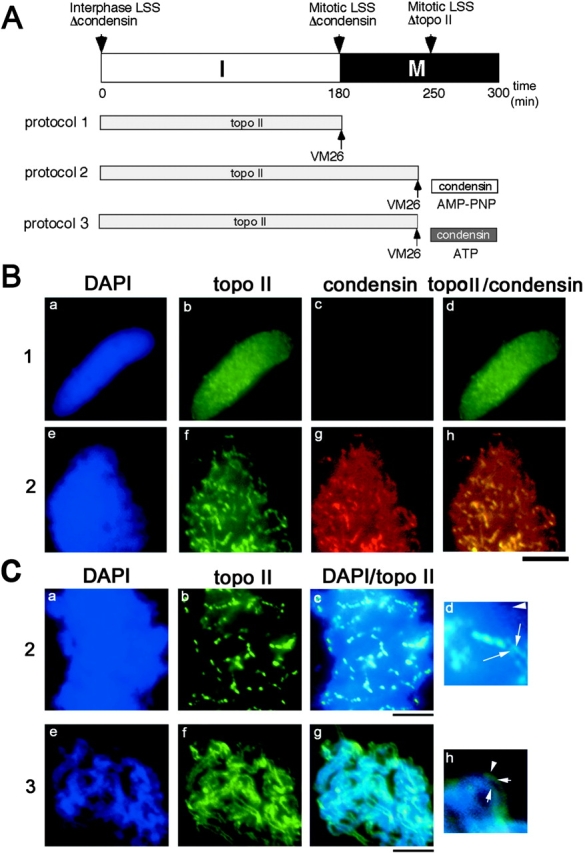
The condensin complex and topoisomerase II (topo II) have different biochemical activities in vitro, and both are required for mitotic chromosome condensation. We have used Xenopus egg extracts to investigate the functional interplay between condensin and topo II in chromosome condensation. When unreplicated chromatin is directly converted into chromosomes with single chromatids, the two proteins must function together, although they are independently targeted to chromosomes. In contrast, the requirement for topo II is temporarily separable from that of condensin when chromosome assembly is induced after DNA replication. This experimental setting allows us to find that, in the absence of condensin, topo II becomes enriched in an axial structure within uncondensed chromatin. Subsequent addition of condensin converts this structure into mitotic chromosomes in an ATP hydrolysis-dependent manner. Strikingly, preventing DNA replication by the addition of geminin or aphidicolin disturbs the formation of topo II-containing axes and alters the binding property of topo II with chromatin. Our results suggest that topo II plays an important role in an early stage of chromosome condensation, and that this function of topo II is tightly coupled with prior DNA replication.


Figures

Figure 1.
Figure 1.
Condensin and topo II play distinct roles in chromosome assembly. (A) A mitotic HSS was immunodepleted with an anti-topo II (lanes 2 and 13) antiserum or a mixture of affinity-purified condensin antibodies (lanes 8 and 15). Mock-depleted extracts were prepared using nonimmune antiserum (lane 1) or IgG (lanes 7 and 14). Equal volumes of these extracts were subjected to 7.5% SDS-PAGE, blotted onto a nitrocellulose paper, and probed with the anti-topo II (lanes 1–6), a mixture of anti-condensin antibodies (lanes 7–12), or both (lanes 13–16). A dilution series of untreated extract (lanes 3–6 and 9–12) was analyzed in parallel to estimate the efficiency of immunodepletion. (B) Chromosomes were assembled from sperm chromatin in mock-depleted (a, d, and g), condensin-depleted (b, e, and h), or topo II–depleted (c, f, and i) HSS. After assembly, the mixture was treated with the indicated extra concentrations of NaCl for 20 min at 22°C, fixed, and stained with DAPI. Bar, 10 μm. (C) Chromosomes were assembled in mock-depleted (a, b, e, and f) topo II–depleted (c and d) or condensin-depleted (g and h) HSS, and immunostained with anti-condensin (XCAP-G; b and d) or anti-topo II antibody (f and h). DNA was counterstained with DAPI (a, c, e, and g). Bar, 10 μm. (D) Topo II assays. (Left) Supercoiled DNA was incubated in the control or depleted extracts in the presence of an increasing concentration of VM-26. The DNA was purified, fractionated on a 0.7% agarose gel, and stained with EtBr. The positions of circular (C), nicked-circular (NC), linear (L), and supercoiled (SC) DNA are indicated. (Middle) Kinetoplast DNA was incubated with the extracts and the reaction was terminated at the indicated time points. The DNA was deproteinized and analyzed on a 0.8% agarose gel. In the mock- and condensin-depleted extracts, kinetoplast DNA was rapidly decatenated to produce relaxed minicircles that became supercoiled as a result of nucleosome assembly. In the topo II–depleted extract, the catenated DNA remained in the well and was not resolved in the gel. (Right) Sperm chromatin was incubated with the depleted extracts to allow chromosome assembly. An increasing concentration of VM-26 (as indicated) was then added to induce topo II–mediated DNA cleavage.
Figure 2.
Figure 2.
Topo II and condensin must function simultaneously to assemble chromosomes in the absence of preceding DNA replication. (A) Experimental scheme. Sperm chromatin was incubated with interphase HSS that had been immunodepleted of condensin. After incubation at 22°C for 180 min, the assembly mixture was converted into mitosis by adding condensin-depleted mitotic HSS. After another 60 min (at 240 min), the mixture was split into two aliquots; one was treated with no drug (protocol 1), and the other was treated with VM-26 at a final concentration of 10 μM (protocol 2). At 250 min, topo II–depleted mitotic HSS was added to allow condensin to function. (B) Chromosomes were assembled according to protocol 1 (a–d) or protocol 2 (e–h), fixed at 300 min, and stained with anti-condensin (XCAP-G; b and f) and anti-topo II antibodies (c and g). DNA was counterstained with DAPI (a and e), and DNA replication was assayed with incorporation of biotinylated dUTP (d and h). Bar, 10 μm.
Figure 3.
Figure 3.
Temporal requirements for topo II and condensin are separable when chromosome assembly is induced after DNA replication. (A) Essentially the same procedure was used as in Fig. 2 A, except that LSS were used instead of HSS. No VM-26 was added in protocol 1. VM-26 was added at 240 min (60 min after mitotic entry) in protocol 2, or at 180 min (at the onset of mitosis) in protocol 3. (B) Chromosomes were assembled according to protocol 1 (a–d), protocol 2 (e–h), or protocol 3 (i–l), fixed at 300 min, and stained with anti-condensin (XCAP-G; b, f, and j) and anti-topo II antibodies (c, g, and k). DNA was counterstained with DAPI (a, e, and i), and DNA replication was assayed with incorporation of biotinylated dUTP (d, h, and l). Bar, 10 μm.
Figure 4.
Figure 4.
Topo II is concentrated on axial structures in mitosis whose assembly depends on preceding DNA replication. (A) Sperm chromatin was incubated with interphase LSS that had been immunodepleted of condensin. After incubation at 22°C for 180 min, interphase chromatin (a–d) was converted into mitosis by adding either condensin-depleted mitotic LSS (e–h and m–p) or purified recombinant cyclin BΔ90 (cyc Δ90; i–l). When required, VM-26 was added before mitotic entry (m–p). Staining was done with anti-condensin (XCAP-G; b, f, j, and n) and anti-topo II (c, g, k, and o) antibodies. DNA was counterstained with DAPI (a, e, i, and m), and DNA replication was assayed with incorporation of biotinylated dUTP (d, h, l, and p). Insets in the third column represent higher magnification of the topo II signals. Bar, 10 μm (3.3 μm for the insets). More examples of topo II–containing axes are also shown below (q–s). The arrows show two paralleled axes stained with anti-topo II. Bar, 1 μm. (B) Sperm chromatin was incubated with interphase LSS that had been immunodepleted of condensin in the presence of aphidicolin (e–h), geminin (i–l), or neither of them (a–d). After incubation for 180 min, cyclin BΔ90 was added to induce mitosis. The assembled structures were stained as in A. Insets in the third column represent higher magnification of the topo II signals. The arrows show two paralleled axes stained by topo II. Bar, 10 μm (3.3 μm for the insets).
Figure 5.
Figure 5.
Topo II–containing axes can be converted by condensin into chromosomes. (A) Experimental scheme. Sperm chromatin was incubated with interphase that had been immunodepleted of condensin. After incubation at 22°C for 180 min (protocol 1), interphase chromatin (a–d) was converted into mitosis by adding condensin-depleted mitotic LSS. After another 60 min (at 240 min), the mixture was split into two aliquots; one was treated with 10 μM VM-26 and 1 mM AMP-PNP (protocol 2), and the other was treated with 10 μM VM-26 with 1mM ATP (protocol 3). At 250 min, topo II–depleted mitotic LSS was added to allow condensin to function. (B) Interphase chromatin was either fixed at 180 min (a–d) or converted to mitosis (e–h) after protocol 1 or 2, respectively. After chromosomes were assembled, staining was done with anti-topo II (b and f) and anti-condensin (XCAP-G; c and g). The merged images of topo II and condensin are also shown (d and h). DNA was counterstained with DAPI (a and e). Bar, 10 μm. (C) Chromosomes were assembled after protocol 2 (a–d) or 3 (e–h), and staining was done with anti-topo II (b and f). DNA was counterstained with DAPI (a and e). The merged images of topo II and DNA are also shown (c and g). Insets (d and h) represent higher magnification of the topo II–containing axes (arrows) and DNA region (arrowheads). Bar, 10 μm (for a–c and e–g); 2 μm (for d and h).
Figure 6.
Figure 6.
DNA replication allows topo II to be tightly associated with chromosomes. (A) Sperm chromatin was incubated with interphase LSS, and then half a volume of mitotic LSS was added to assemble double-chromatid chromosomes (a–h). Alternatively, a combination of interphase and mitotic HSS was used to assemble single-chromatid chromosomes without DNA replication (i–p). The assembly mixtures were untreated (a–d and i–l) or treated with 0.6 M extra concentrations of NaCl (e–h and m–p) for 20 min at 22°C, fixed, and stained with anti-topo II (b, f, j, and n), anti-condensin (XCAP-G; c, g, k, and o), and DAPI (a, e, i, and m). DNA replication was assayed with incorporation of biotinylated dUTP (d, h, l, and p). Bar, 1 μm. (B) Chromosomes were assembled in LSS in the presence of aphidicolin, and the assembly mixtures were untreated (a–d) or treated as above with 0.6 M NaCl (e–h). (C) Chromosomes were assembled in LSS that had been immunodepleted of condensin, and the mixture was untreated (a–d) or treated as above with 0.6 M NaCl (e–h). Staining was performed as described in A. Bar, 1 μm.
Figure 7.
Figure 7.
Chromosome condensation in the presence or absence of DNA replication. For details, see the text.

Similar articles

Cited by

References

    1. Adachi, Y., M. Luke, and U.K. Laemmli. 1991. Chromosome assembly in vitro: topoisomerase II is required for condensation. Cell. 64:137–148. - PubMed
    1. Anderson, D.E., A. Losada, H.P. Erickson, and T. Hirano. 2002. Condensin and cohesin display different arm conformations with characteristic hinge angles. J. Cell Biol. 156:419–424. - PMC - PubMed
    1. Bazzett-Jones, D.P., K. Kimura, and T. Hirano. 2002. Efficient supercoiling of DNA by a single condensin complex as revealed by electron epectroscopic imaging. Mol. Cell. 9:1183–1190. - PubMed
    1. Bhalla, N., S. Biggins, and A.W. Murray. 2002. Mutation of YCS4, a budding yeast condensin subunit, affects mitotic and nonmitotic chromosome behavior. Mol. Biol. Cell. 13:632–645. - PMC - PubMed
    1. Bhat, M.A., A.V. Philp, D.M. Glover, and H.J. Bellen. 1996. Chromatid segregation at anaphase requires the barren product, a novel chromosome associated protein that interacts with topoisomerase II. Cell. 87:1103–1114. - PubMed

Publication types

MeSH terms