. 2003 Mar 4;100(5):2462-7.
doi: 10.1073/pnas.0437964100. Epub 2003 Feb 25.

Nonhomologous end joining and V(D)J recombination require an additional factor

Affiliations

Nonhomologous end joining and V(D)J recombination require an additional factor

Y Dai et al. Proc Natl Acad Sci U S A. .

Abstract

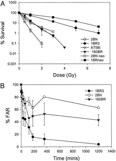
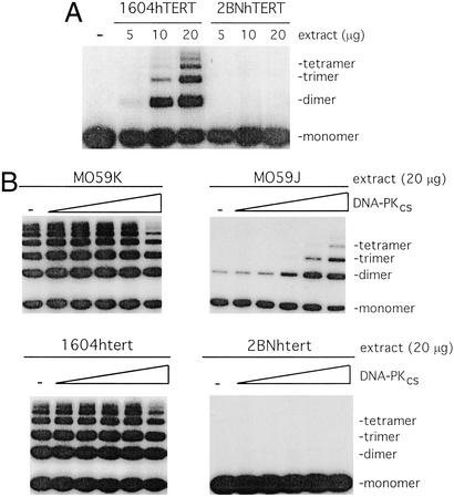
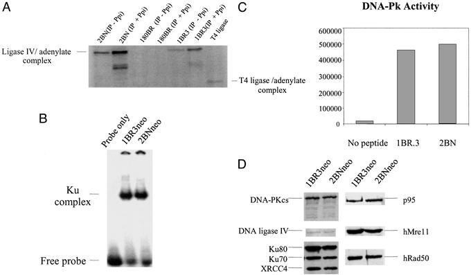
DNA nonhomologous end-joining (NHEJ) is the major pathway for repairing DNA double-strand breaks in mammalian cells. It also functions to carry out rearrangements at the specialized breaks introduced during V(D)J recombination. Here, we describe a patient with T(-)B(-) severe combined immunodeficiency, whose cells have defects closely resembling those of NHEJ-defective rodent cells. Cells derived from this patient show dramatic radiosensitivity, decreased double-strand break rejoining, and reduced fidelity in signal and coding joint formation during V(D)J recombination. Detailed examination indicates that the patient is defective neither in the known factors involved in NHEJ in mammals (Ku70, Ku80, DNA-dependent protein kinase catalytic subunit, Xrcc4, DNA ligase IV, or Artemis) nor in the Mre11/Rad50/Nbs1 complex, whose homologue in Saccharomyces cerevisiae functions in NHEJ. These results provide strong evidence that additional activities are crucial for NHEJ and V(D)J recombination in mammals.


Figures

Figure 1
Figure 1
2BN cells show radiosensitivity and defective DSB repair. (A) Survival of 2BN cells after exposure to IR. Cells were exposed to γ-rays and survival was estimated by colony formation as described (12). The data represent the mean results of a minimum of three experiments for each cell line, and error bars represent the standard deviation of the mean. The survival response of 2BNneo cells represents the mean of two experiments, and error bars have not been included. The survival of 2BNhTERT cells (the mean of two experiments) was identical to that of 2BN primary cells. (B) DSB repair in 2BN cells; estimation of DNA DSB rejoining by PFGE. Cells, radiolabeled with [14C]-TdR, were embedded in agarose, irradiated with 40 Gy γ-rays, incubated for varying times, and subjected to PFGE. The fraction of activity released (FAR) represents the radioactivity that enters the gel, divided by the total activity (well + lane). The results are presented as the ratio of the FAR remaining at the specified time compared with the FAR at time 0. The FAR for unirradiated cells is subtracted before these estimations.
Figure 2
Figure 2
2BNhTERT cells fail to promote end-joining in vitro. (A) Cell-free extracts prepared from either the control cell line 1604hTERT or 2BNhTERT were incubated with 5′-32P-end-labeled linear plasmid DNA. The products of end-joining were analyzed by gel electrophoresis. (B) Extracts from either 1604hTERT, 2BNhTERT, DNA-PKcs-proficient cells (MO59K), or DNA-PKcs defective cells (MO59J) were incubated with 5′-32P-end-labeled linear plasmid DNA. Where indicated, extracts were complemented by addition of purified DNA-PKcs (0, 0.14, 0.43, 1.3, 3.9, or 11.6 nM). DNA products were analyzed as described in A.
Figure 3
Figure 3
2BN cells show normal expression and activity of characterized NHEJ proteins. (A) DNA ligase IV adenylation activity. Because DNA ligase IV antibodies are inefficient in IP, anti-Xrcc4 antibodies were used to coimmunoprecipitate DNA ligase IV. DNA ligase IV exists endogenously as an enzyme–adenylate complex; therefore, extracts were or were not treated with 5 mM inorganic pyrophosphate (Ppi) to disrupt preformed AMP–ligase complexes and then analyzed for DNA ligase IV–adenylate complex formation. As expected, the signal is greater after Ppi treatment. DNA ligase IV-defective 180BR cells were included as a control (21). (B) DNA end-binding activity. Whole-cell extracts (5 μg) from 1BR3 and 2BN cells were incubated with [γ-32P]dATP-labeled M1/M2 oligonucleotide probe and separated by PAGE. The position of the free probe and Ku-dependent bands is highlighted. The band was shown to be Ku-dependent, because it is absent in Ku80-defective Xrs-6 cells and is regained in xrs-6 complemented with human Ku80 cDNA (data not shown and ref. 22). (C) DNA-Pk activity. DNA-binding proteins were microfractionated from whole-cell extracts (10 μg) of 1BR3 and 2BN cells using DNA cellulose beads and incubated with a p53-derived peptide in the presence of [γ-32P]dATP. The peptide was separated from other nonspecific phosphorylated proteins by PAGE and quantified by PhosphorImager counting. The results represent arbitrary PhosphorImager units. (D) Western blot analysis of NHEJ proteins in 2BN cells. Whole-cell extracts of transformed 2BN and control cells were analyzed by Western blotting by using antibodies against DNA ligase IV, Ku80 (Ku80-4), Ku70 (N3H10), Xrcc4, and DNA-PKcs (MSAG3). Anti-hRad50, hMre11, and p95 antibodies were a kind gift from J. H. J. Petrini (35).

References

    1. Taccioli G E, Rathbun G, Oltz E, Stamato T, Jeggo P A, Alt F W. Science. 1993;260:207–210. - PubMed
    1. Nussenzweig A, Chen C, da Costa Soares V, Sanchez M, Sokol K, Nussenzweig M C, Li G C. Nature. 1996;382:551–555. - PubMed
    1. Fugmann S D, Lee A I, Shockett P E, Villey I J, Schatz D G. Annu Rev Immunol. 2000;18:495–527. - PubMed
    1. Gellert M. Adv Immunol. 1997;64:39–64. - PubMed
    1. Jeggo P A. In: Advances in Genetics. Hall J C, Dunlap J C, Friedmann T, Giannelli F, editors. Vol. 38. San Diego: Academic; 1998. pp. 185–211.

Publication types

MeSH terms