. 2003 Mar 4;100(5):2450-5.
doi: 10.1073/pnas.0437902100. Epub 2003 Feb 25.

Disorder in the nuclear pore complex: the FG repeat regions of nucleoporins are natively unfolded

Affiliations

Disorder in the nuclear pore complex: the FG repeat regions of nucleoporins are natively unfolded

Daniel P Denning et al. Proc Natl Acad Sci U S A. .

Abstract

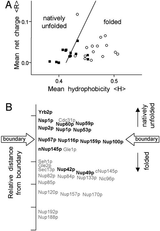
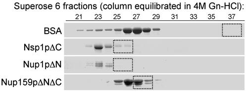
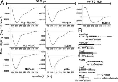
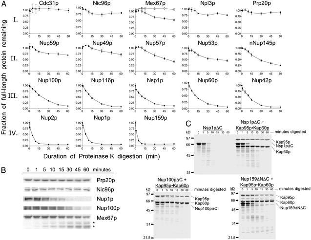
Nuclear transport proceeds through nuclear pore complexes (NPCs) that are embedded in the nuclear envelope of eukaryotic cells. The Saccharomyces cerevisiae NPC is comprised of 30 nucleoporins (Nups), 13 of which contain phenylalanine-glycine repeats (FG Nups) that bind karyopherins and facilitate the transport of karyopherin-cargo complexes. Here, we characterize the structural properties of S. cerevisiae FG Nups by using biophysical methods and predictive amino acid sequence analyses. We find that FG Nups, particularly the large FG repeat regions, exhibit structural characteristics typical of "natively unfolded" proteins (highly flexible proteins that lack ordered secondary structure). Furthermore, we use protease sensitivity assays to demonstrate that most FG Nups are disordered in situ within the NPCs of purified yeast nuclei. The conclusion that FG Nups constitute a family of natively unfolded proteins supports the hypothesis that the FG repeat regions of Nups form a meshwork of random coils at the NPC through which nuclear transport proceeds.


Figures

Figure 1
Figure 1
Computer analysis of the hydrophobicity and net charge of all S. cerevisiae Nups predicts that the FG Nups are unfolded in physiological conditions. (A) Charge versus hydrophobicity plot. The mean net charges and mean hydrophobicities (Kyte-Doolittle, 5-aa window) were calculated and plotted for FG Nups (■) and non-FG Nups (○) as described (11). The line, 〈H〉b = (〈R〉 + 1.151)/2.785, demarcates the observed boundary between folded and natively unfolded proteins in the charge-hydrophobicity plot. The relative distance to the boundary line for individual FG Nups (bold type) and non-FG Nups (gray type) is shown on a linear axis in B.
Figure 2
Figure 2
Elution profiles of FG Nups in a Superose 6 gel filtration column are similar in the presence and absence of 4 M Gn-HCl. Purified FG Nups and BSA (≈100 μg each) were separated by size in a Superose-6 column (24 ml) equilibrated with 4 M Gn-HCl; 0.5-ml fractions were collected, and the eluted proteins were precipitated with trichloroacetic acid, separated by SDS/PAGE, and stained with Coomassie blue. Boxes identify the elution peak of the protein separated in the same Superose 6 column equilibrated with physiological buffer [20 mM Hepes, pH 6.8/150 mM KOAc/2 mM Mg(OAc)2].
Figure 3
Figure 3
(A) Far-UV CD measurements of purified Nups or Nup fragments show extensive regions of structural disorder in FG Nups. Shown are the CD spectra of purified recombinant proteins. The portion of each Nup used is noted by brackets in B. All spectra were obtained at 25°C at neutral pH in 1-mm cuvettes as described (10). Protein concentrations ranged from 0.05 to 0.2 mg/ml. (B) Diagram of FG Nups analyzed. The portion of each FG Nup that was purified and analyzed is bracketed; in the case of Yrb2p and Nup2p, the full-length protein was characterized. The content of disordered structure as determined by FTIR analysis (see Table 3) is given as a percentage of the fragment analyzed.
Figure 4
Figure 4
FG Nups within NPCs in purified nuclei are hypersensitive to protease digestion. (A) Purified nuclei (4 mg/ml) were digested with proteinase K (300 ng/ml) for the indicated times at 37°C. Aliquots were mixed with SDS buffer with 2 mM PMSF to stop the digestion. The degradation of specific proteins was quantified by using Western blots probed with specific antibodies and 125I-Protein A. For each protein, the fraction of full-length protein remaining is shown. Each graph represents the average of three to six independent digestions; error bars represent the SEM. The top line in Mex67p represents the sum of full-length and clipped Mex67p (*); the bottom line represents full-length Mex67p. (B) Representative 125I-Protein A Western blots used to generate graphs shown in A. Note that in the case of Mex67p, the full-length protein is clipped to yield two large, stable fragments (*). (C) Purified FG repeat regions of Nups (1 mg/ml) with and without stoichiometric amounts of Kap95p–Kap60p were incubated with proteinase K (100 ng/ml) for the indicated times at 37°C. Samples were processed as in A, resolved by SDS/PAGE, and visualized by Coomassie blue staining.

References

    1. Rout M, Aitchison J. J Biol Chem. 2001;276:16593–16596. - PubMed
    1. Yang Q, Rout M P, Akey C W. Mol Cell. 1998;1:223–234. - PubMed
    1. Rexach M, Blobel G. Cell. 1995;83:683–692. - PubMed
    1. Allen N, Huang L, Burlingame A, Rexach M. J Biol Chem. 2001;276:29268–29274. - PubMed
    1. Gorlich D, Kutay U. Annu Rev Cell Dev Biol. 1999;15:607–660. - PubMed

MeSH terms