. 2003 Mar 4;100(5):2706-11.
doi: 10.1073/pnas.0436037100. Epub 2003 Feb 25.

von Hippel-Lindau protein binds hyperphosphorylated large subunit of RNA polymerase II through a proline hydroxylation motif and targets it for ubiquitination

Affiliations

von Hippel-Lindau protein binds hyperphosphorylated large subunit of RNA polymerase II through a proline hydroxylation motif and targets it for ubiquitination

Anna V Kuznetsova et al. Proc Natl Acad Sci U S A. .

Abstract

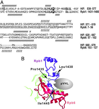
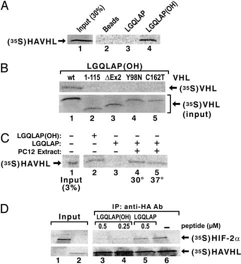
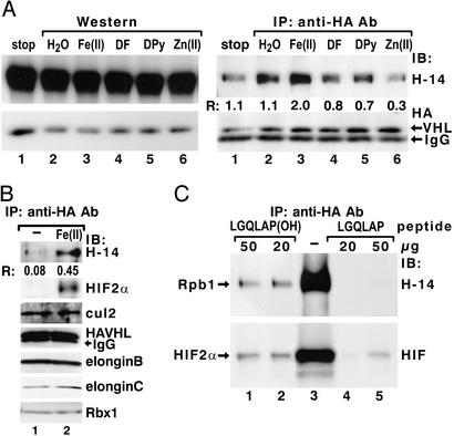
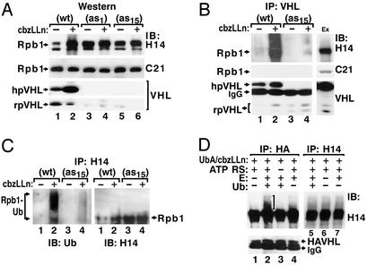
The transition from transcription initiation to elongation involves phosphorylation of the large subunit (Rpb1) of RNA polymerase II on the repetitive carboxyl-terminal domain. The elongating hyperphosphorylated Rpb1 is subject to ubiquitination, particularly in response to UV radiation and DNA-damaging agents. By using computer modeling, we identified regions of Rpb1 and the adjacent subunit 6 of RNA polymerase II (Rpb6) that share sequence and structural similarity with the domain of hypoxia-inducible transcription factor 1 alpha (HIF-1 alpha) that binds von Hippel-Lindau tumor suppressor protein (pVHL). pVHL confers substrate specificity to the E3 ligase complex, which ubiquitinates HIF-alpha and targets it for proteasomal degradation. In agreement with the computational model, we show biochemical evidence that pVHL specifically binds the hyperphosphorylated Rpb1 in a proline-hydroxylation-dependent manner, targeting it for ubiquitination. This interaction is regulated by UV radiation.


Figures

Figure 1
Figure 1
Computational prediction of the pVHL-binding pocket in the RNA polymerase II complex. (A) Sequence-to-structure alignments of the HIF-1α ODDD fragment into the carboxyl-terminal fragments of human Rpb1 and Rpb6 subunits. The Rpb1 and Rpb6 secondary structures are indicated below, and the predicted HIF-1α secondary structures are shown above their sequences. H, α (and other)-helices; E, extended β-strands. HIF-1α motifs that make contact with the pVHL complex (including the Pro-564 residue) are shaded and, if conserved in Rpb1, bold. The critical HIF-1α residues (L559, L562, P564, and D571) are conserved in the Rpb1 structure. The K532 residue, ubiquitinated on HIF-1α, is boxed. The human and yeast Rpb6 structures (PDB ID codes 1QKL and 1I50, chain F, respectively) are different by an additional β-strand occurring only on the human Rpb6 structure (boxed fragment). (B) The predicted pVHL-binding pocket (Rpb1, purple; Rpb6, red; other fragments in contact with the binding pocket are green). The critical proline residue and the flanking amino acids are indicated by using ball and stick models of their side chains. The numbering of residues is according to the yeast Rpb1 structure with the yeast Leu-1430, Pro-1435, and Ile-1445 residues corresponding to Leu-1460, Pro-1465, and Leu-1475 of the human Rpb1, respectively.
Figure 2
Figure 2
pVHL binds to the Rpb1 synthetic 36-aa peptide with hydroxylated P1465. (A) Binding of [35S]pVHL to the Rpb1 peptide hydroxylated (lane 4), or nonhydroxylated (lane 3), on P1465. (B) Binding of the in vitro-translated mutated forms of pVHL to the hydroxylated peptide. To ensure that the amounts of the labeled mutant proteins used in the peptide-binding reactions were the same as for the WT pVHL, the amounts of the lysate with radioactively labeled mutant proteins used in the binding reactions were normalized accordingly by using the PhosphorImager quantification. (C) Hydroxylation of the Rpb1 peptide in extract from PC12 cells. HAVHL, HA-tagged pVHL. (D) Competition experiment of [35S]HIF-2α–[35S]pVHL binding by hydroxylated (lanes 3 and 4) or nonhydroxylated (lane 5) peptide.
Figure 3
Figure 3
pVHL specifically interacts with the hyperphosphorylated Rpb1 in nuclear extracts from PC12 and RCC cells. Coimmunoprecipitations (IP) using monoclonal antibodies against HA or pVHL or mouse anti-rabbit IgG (RG-96) in nuclear extracts from PC12 cells overexpressing human HA-tagged pVHL [PC12 VHL (WT)] (A and B), or control PC12 cells stably transfected with an empty vector (A). (C) Dephosphorylation of Rpb1 in PC12 cellular extracts for the indicated times by treating the extracts with alkaline phosphatase (AP) in the absence (lanes 3, 4, 7, and 8) or presence (lanes 6 and 9) of NaF. Treated extracts were subjected to immunoprecipitations using anti-HA antibodies. (D) Anti-HA immunoprecipitations in nuclear extracts from RCC 786-O cells lacking pVHL function (lanes 1 and 2) or from cells stably transfected with HA-pVHL (1) (lanes 3 and 4). PC12 and indicated RCC cells were pretreated with 10 μM CbzLLn for 6 h to increase accumulation of the hyperphosphorylated Rpb1. The immunoprecipitates were washed with high-detergent immunoprecipitation buffer (A, C, and D), or immunoprecipitation buffers containing up to 900 mM NaCl and 0.5% Igepal (B). Blots were probed with the indicated antibodies, human (h)pVHL and rat (r)pVHL, respectively.
Figure 4
Figure 4
pVHL binds Rpb1 in a proline hydroxylation-dependent manner. (A) Preincubation of PC12 cellular extracts with FeCl2, ascorbic acid, and 2-oxoglutarate (100 μM each) (lane 3), or with 100 μM each of iron chelators: desferrioxamine (DF, lane 4) and 2,2′-dipyridyl (DPy, lane 5), or ZnCl2 (lane 6), followed by Western blot analysis (Left) or coimmunoprecipitations (IP) with anti-HA antibodies (Right). IB, immunoblotting antibody. (B) Coimmunoprecipitation of the components of pVHL-associated complex by using anti-HA antibody in cellular lysates (lane 1) or lysates treated under hydroxylating conditions with Fe(II), ascorbate, and 2-oxoglutarate, as in A. Immunoblots were probed with the indicated antibodies. (C) Elution of hyperphosphorylated Rpb1 and HIF-2α with a hydroxylated 36-aa Rpb1 peptide. R describes the ratio of the signal detected with H14 antibody to the signal detected with anti-HA antibody, as quantified by using optical density measurements.
Figure 5
Figure 5
Accumulation and ubiquitination of hyperphosphorylated Rpb1 in cells with different levels of pVHL. (A) Western blot analysis of hyperphosphorylated (H14) and hypophosphorylated (C21) Rpb1 in nuclear extracts from PC12VHL(WT) or two different clones of PC12VHL antisense (as) cells. (B) Coimmunoprecipitations using anti-pVHL antibody from nuclear extracts of WT and antisense cells. Ex, extract. (C) Immunoprecipitation of ubiquitinated forms of hyperphosphorylated Rpb1 from denatured cellular lysates by using H14 antibody. (D) In vitro ubiquitination reactions on protein complexes coimmunoprecipitated by using anti-HA (lanes 1–4) or H14 (lanes 5–7) antibodies from cellular extracts from PC12VHL(WT) cells. H14 antibody does not coimmunoprecipitate pVHL. UbA, ubiquitin aldehyde; E, purified enzymatic fraction II from reticulocyte lysate; ATP RS, ATP-regenerating solution. The bracket marks ubiquitinated forms of Rpb1.
Figure 6
Figure 6
Rpb1–pVHL interactions in response to the UV treatment. (A) Western blot analysis of Rpb1 and pVHL in nuclear extracts from cells in the absence (Upper) or presence (Lower) of CbzLLn. (B) Coimmunoprecipitation of hyperphosphorylated Rpb1 with anti-HA antibody in nuclear extracts from cells treated with UV for the indicated times. Blots were probed with indicated antibodies. (C) Ubiquitination of the hyperphosphorylated Rpb1 in response to the UV treatment in PC12VHL(WT) and antisense cells. Two hours after UV irradiations denatured cellular lysates were immunoprecipitated with H14 antibodies and the immunoblots were probed with anti-ubiquitin antibody.

References

    1. Kibel A, Iliopoulos O, DeCaprio J A, Kaelin W G., Jr Science. 1995;269:1444–1446. - PubMed
    1. Pause A, Lee S, Worrell R A, Chen D Y, Burgess W H, Linehan W M, Klausner R D. Proc Natl Acad Sci USA. 1997;94:2156–2161. - PMC - PubMed
    1. Kamura T, Koepp D M, Conrad M N, Skowyra D, Moreland R J, Iliopoulos O, Lane W S, Kaelin W G, Jr, Elledge S J, Conaway R C, et al. Science. 1999;284:657–661. - PubMed
    1. Cockman M E, Masson N, Mole D R, Jaakola P, Chang G-W, Clifford S C, Maher E R, Pugh C W, Ratcliffe P J, Maxwell P H. J Biol Chem. 2000;275:25733–25741. - PubMed
    1. Kamura T, Sato S, Iwai K, Czyzyk-Krzeska M F, Conaway R C, Conaway J W. Proc Natl Acad Sci USA. 2000;97:10430–10435. - PMC - PubMed

Publication types

MeSH terms